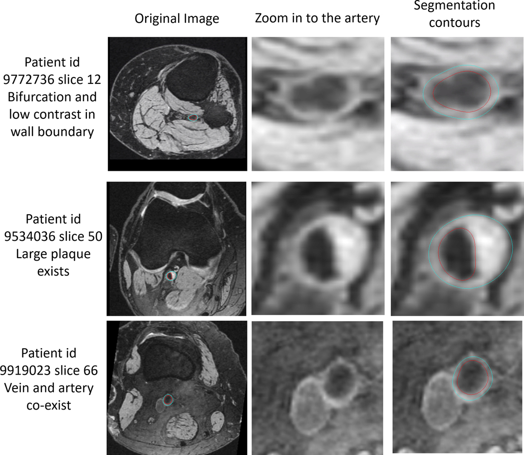
FIGURE 4.
Example of FRAPPE generated contours (right column images: red contour, lumen boundary; blue contour, outer wall boundary) in challenging images (original images shown in the left column, zoomed-in images shown in the middle column) with low image contrast around vessel wall boundary, bifurcation (top row), plaque (middle row), and when artery is close to the vein (bottom row)

