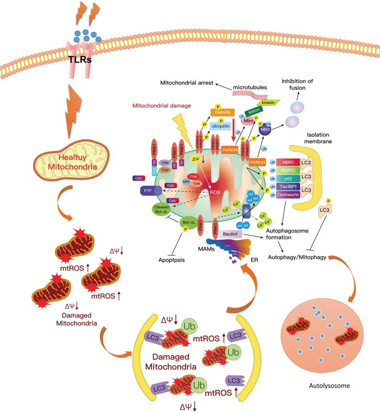
FIGURE 4.
PINK1 could phosphorylate Parkin on the mitochondrial surface, resulting in the activation of Parkin. The activated Parkin proteins form phospho-polyubiquitin chains on damaged mitochondria. Finally, the dysfunctional mitochondria are cleared via autophagy. In this way, mitophagy inhibits neuroinflammation in PD and increases microglial phagocytosis. However, the mutation of PINK1 or Parkin would alter the balance of fission to fusion by preventing cells from responding to mitochondrial damage. The expression of ROS and pro-inflammatory factors are increased, which aggravates the development of PD. PD, Parkinson’s disease; ROS, reactive oxygen species.

