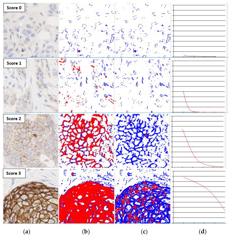
Figure 1.
The shapes of the characteristic curves for images with different human epidermal growth factor receptor 2 (HER2) scores (a) Input image; (b) Thresholded image at saturation 0.1; (c) Thresholded image at saturation 0.5; (d) Characteristic curve with x-axis denoting saturation from 0 to 0.5, and y-axis % of stained region.

