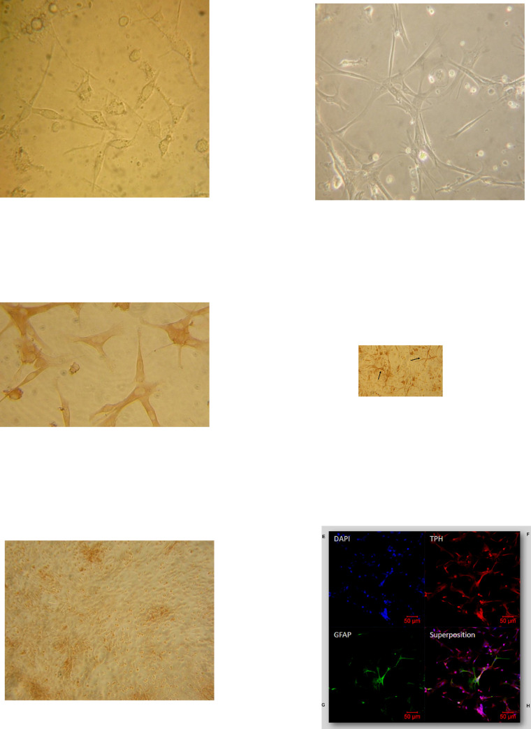
Fig 1. Cytological characterization of primary culture of ovine pineal gland cells.
Five (A) and eight days (B) primary culture of ovine pineal glands were observed by optical microscopy. Cells from ovine pineal glands were immunolabelled with anti-tryptophan hydroxylase (TPH, C) or anti- glial fibrillary acidic protein (GFAP, D) antibodies and detected by a peroxydase-coupled secondary antibody. Non immun serum (NIS) was used as a negative control (Fig 1E). Magnification was x400 in A and C, x200 in B and x100 in D. Pinealocyte were labelled with an anti-TPH antibody (G) detected by a cyanin-coupled secondary antibody, while astrocytes were labelled with an anti-GFAP antibody (H) detected by fluorescein-coupled secondary antibody. Nuclei were stained with DAPI (F). The merge of the three labelings is shown in I. Maginifcation is x10. Scale bar 50 μm. Astrocytes (arrow).

