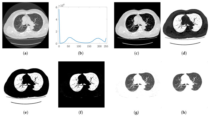
Figure 3.
Automatic histogram based on initial segmentation: (a) Input CT slice; (b) histogram of the image in (a); (c) the results after thresholding the image with the threshold estimated from the histogram in (b); (d) the complemented image of (c); (e) binary segmentation map; (f) map after connected components-based refinement; (g) detected lungs; (h) lungs after noise removal.

