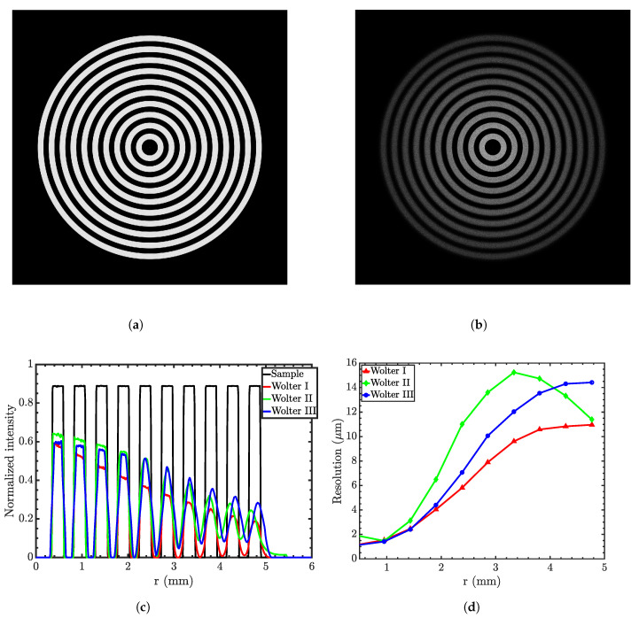
Figure 13.
(a) Concentric rings sample; (b) the magnified image in the Wolter I microscope; (c) ideal edge-spread function (ESF) and that obtained by three types of Wolter mirrors; (d) the resolution at the focal plane as a function of the distance from the optical axis of the three microscopes. Simulations were conducted using 109 rays.

