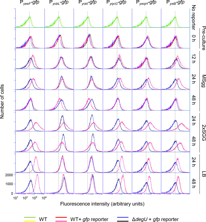
Fig 2. Expression of LXG toxin–antitoxin operons.
Strains harboring promoter-gfp reporters were grown to an OD600 of 0.7 to 0.8 in liquid LB (preculture, 0 h). The cultures were diluted to an OD600 of 0.5, and 2 μl of the dilutions were spotted on three solid media, MSgg, 2×SGG, and LB. After 12 h, 24 h, or 48 h of incubation at 30°C, expression of gfp reporters in colonies were measured by flow cytometry. Strain 3610 (with no gfp reporter) was used as a negative control. Two sets of data are shown for each strain. All histograms are shown on the same scale, and values for the x and y axes are shown only in the lower left histogram. The peak positions of fluorescence from strain 3610 are indicated as a background reference by dotted lines.

