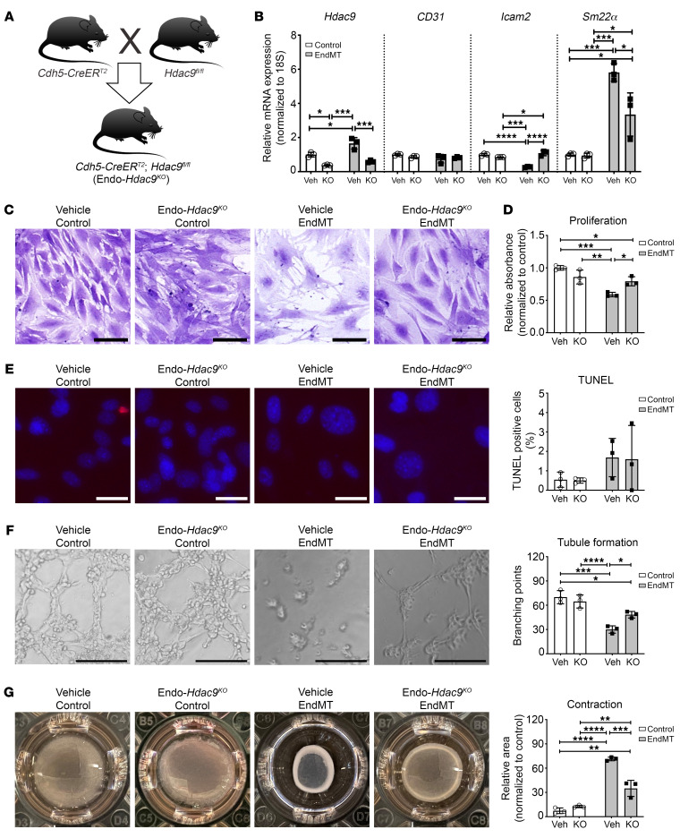
Figure 3. Ex vivo endothelial-specific Hdac9 knockout attenuates EndMT-associated changes in endothelial cell function in MPLECs.
(A) Breeding and generation of Endo-Hdac9KO mice for obtaining MPLECs. (B) Expression of Hdac9, CD31, Icam2, and Sm22α in MPLECs with (KO; i.e., ex vivo 4-OH tamoxifen treatment) and without (Veh; vehicle treatment) knockout of Hdac9 with or without EndMT assessed by qRT-PCR. These groups are identical for all subsequent panels in this figure. (C) Crystal violet staining showing changes in cell numbers and density. Scale bars: 100 μm. (D) To assess proliferation, MPLECs were incubated with BrdU, followed by spectrophotometric quantification. Data are represented as fold change compared with vehicle-treated control cells. (E) Representative images and quantification of TUNEL assay to detect apoptosis on MPLECs with or without EndMT induction. Scale bars: 30 μm. (F) Tubule formation of MPLECs with or without EndMT assessed by plating cells onto Matrigel and incubating for another 4 hours. Tubule branch points were imaged and quantified. Scale bars: 100 μm. (G) Contraction assay showing changes in relative unoccupied area (normalized to a completely unoccupied well) for MPLECs with or without EndMT induction. For this figure, lungs from n = 4 male Endo-Hdac9KO mice were pooled to derive MPLECs. Apart from (crystal violet staining) (C), n = 3 for all analyses as biological replicates. *P ≤ 0.05; **P ≤ 0.01; ***P ≤ 0.001; ****P ≤ 0.0001. All analyses performed using 2-way ANOVA.

