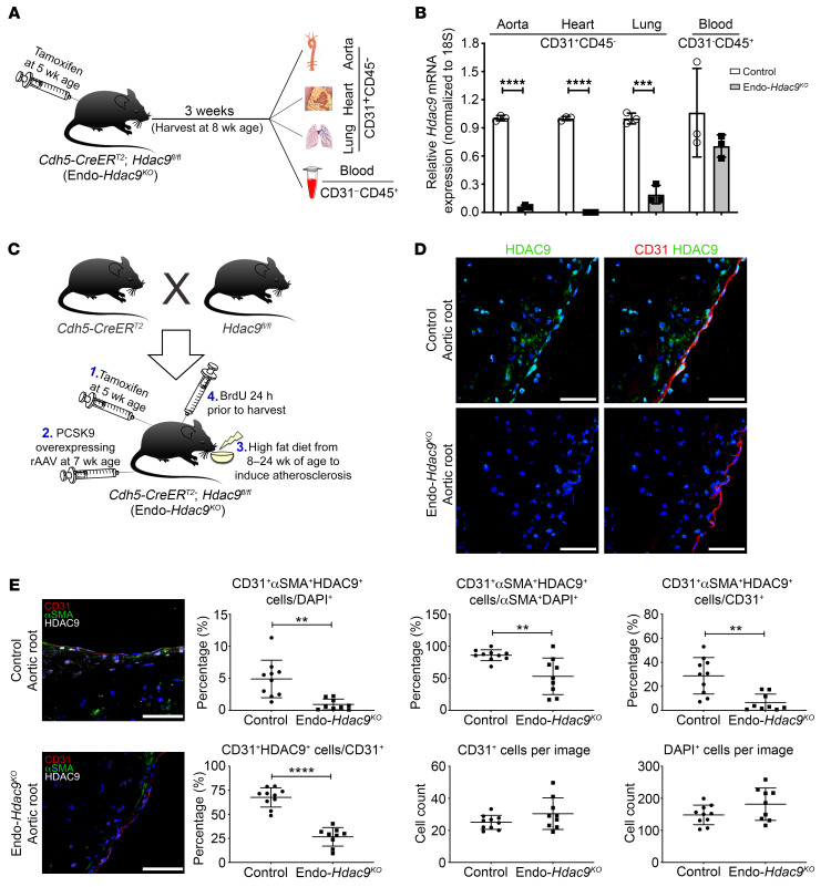
Figure 4. In vivo establishment and validation of Endo-Hdac9KO mouse model.
All comparisons in this figure are using endothelial-specific Hdac9 knockout mice (Endo-Hdac9KO) versus littermate controls (Hdac9fl/fl). All mice received tamoxifen. (A) For Hdac9 knockout validation, endothelial cells were harvested from a variety of tissues from nonatherosclerotic Endo-Hdac9KO mice or littermate controls. (B) Hdac9 knockout validation: qRT-PCR analysis of the expression levels of Hdac9 in CD31+CD45– endothelial cells from the aorta, heart, and lungs and CD31–CD45+ leukocytes from blood in Endo-Hdac9KO mice compared with littermate controls 3 weeks after tamoxifen administration. n = 3. (C) Breeding and generation of atherosclerotic Endo-Hdac9KO mouse model. (D) Representative immunofluorescence staining images for HDAC9- (green), CD31- (red), and DAPI-stained nuclei (blue) in plaques from the aortic root. (E) Representative immunofluorescence staining images for αSMA- (green), CD31- (red), HDAC9- (white), and DAPI-stained nuclei (blue) in aortic root plaques and quantification. Scale bars: 50 μm. n = 10 controls versus n = 9 Endo-Hdac9KO mice. **P ≤ 0.01; ***P ≤ 0.001; ****P ≤ 0.0001. All analyses performed using unpaired Student’s t test except CD31+αSMA+HDAC9+ cells/CD31+ (E), for which Mann-Whitney U test was used.

