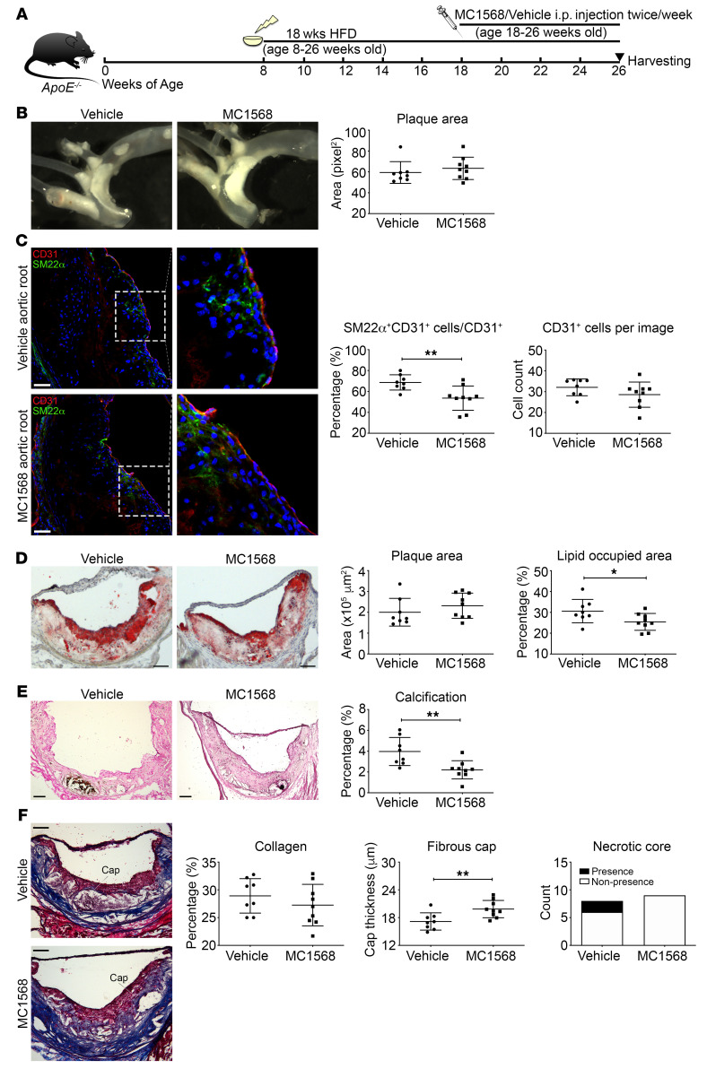
Figure 7. Systemic administration of a class IIa HDAC inhibitor reduces EndMT and increases plaque stability.
(A) Generation of MC1568-treated atherosclerotic mouse model. (B) Representative images of aorta before embedding into OCT and en face analysis of plaque in aorta. (C) Representative images of immunofluorescence staining of aortic root sections for SM22α- (green), CD31- (red), and DAPI-stained nuclei (blue) and quantification of SM22α+CD31+-costained cells over total CD31+ cells and the total number of CD31+ cells per image. (D) Representative images of the aortic root with staining using oil red O, with quantification of total plaque area and plaque lipid content. (E) Representative images of aortic root sections using Von Kossa stain (black represents calcification), with quantification of calcification. (F) Representative aortic root images stained with Masson’s trichrome with quantification of collagen content, fibrous cap thickness, and presence/nonpresence of necrotic core (blue, collagen; pink, macrophages and cardiac muscle). Scale bars: 50 μm (C); 100 μm (D–F). Right panels in C are digital enlargements of the original adjacent images. n = 8 controls (vehicle) versus n = 9 MC1568-treated mice for all panels. *P ≤ 0.05; **P ≤ 0.01. All analyses performed using unpaired Student’s t test except plaque area (B and D), for which Mann-Whitney U test was used. In addition, presence or nonpresence of necrotic core (F) was analyzed using Fisher’s exact test.

