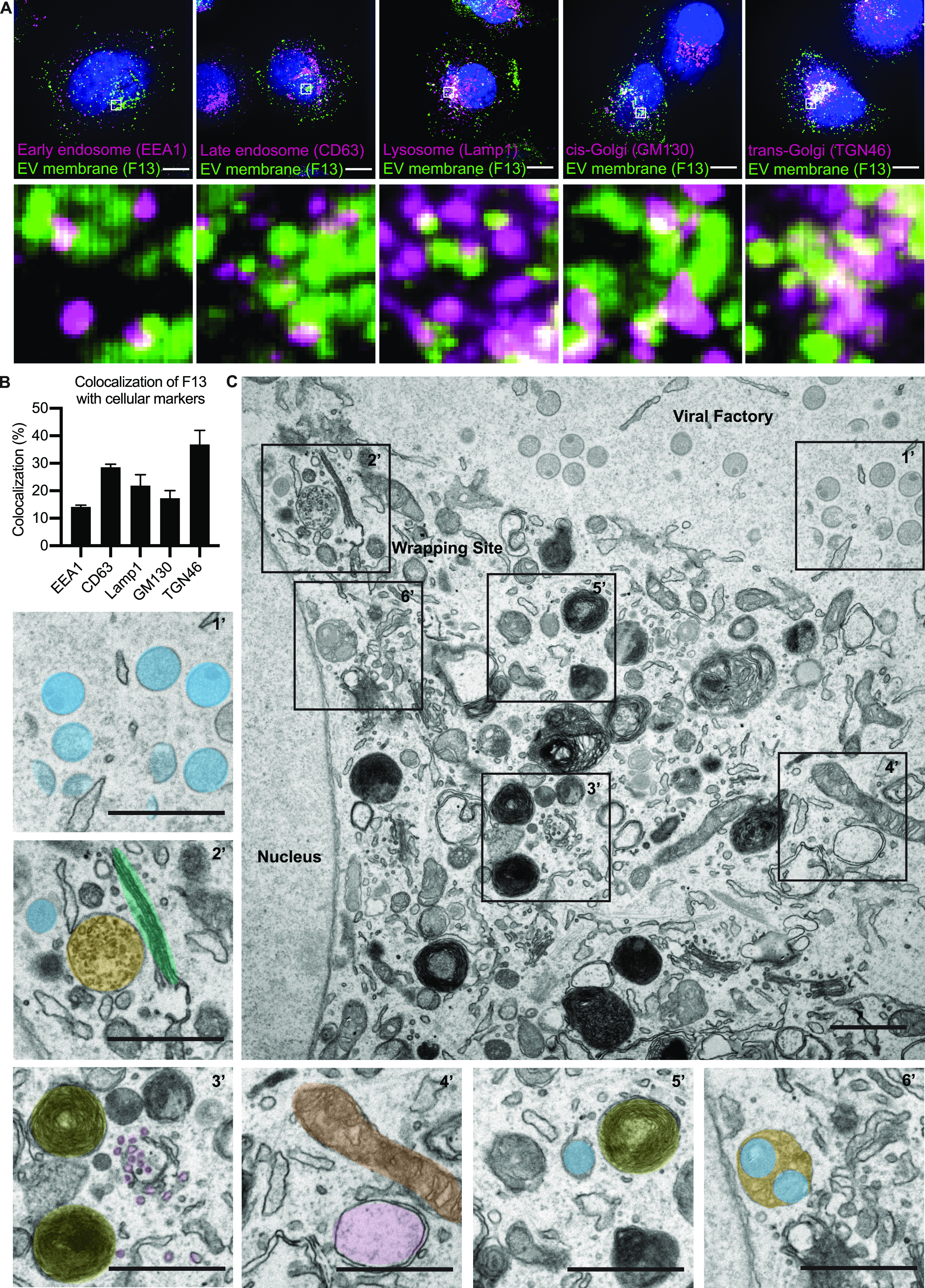
Figure 5. MVBs serve as a second major source of VACV wrapping membrane.

(A) Immunofluorescent (IF) imaging 8 hpi shows several cellular membrane markers in close proximity of EV membrane protein F13 (EEA1, CD63, Lamp1, GM130, and TGN46). Maximum intensity projections. Scale bars = 10 µm. Insets = 20× zoom. (A, B) Quantification of F13 colocalization with cell markers from (A). (C) EM imaging 8 hpi illustrates the location of viral replication site (viral factory) with different stages of mature virion morphogenesis (crescents and immature virions [1’]). In addition, the imaging shows that in the areas of intracellular mature virion wrapping, various cellular membrane structures are in close proximity of wrapping virions. 2’: blue = intracellular mature virion, green = Golgi stacks, orange = multivesicular body (MVB), 3’: yellow = lysosomes, purple = small vesicles, 4’: brown = mitochondria, pink = early endosome, 5’: blue = wrapping virion, yellow = lysosome, 6’: orange/blue = virions bud into MVB. Scale bars = 1 µm.
