
Figure 2. IL-13-dependent airway eosinophilia during Nippostrongylus brasiliensis infection.
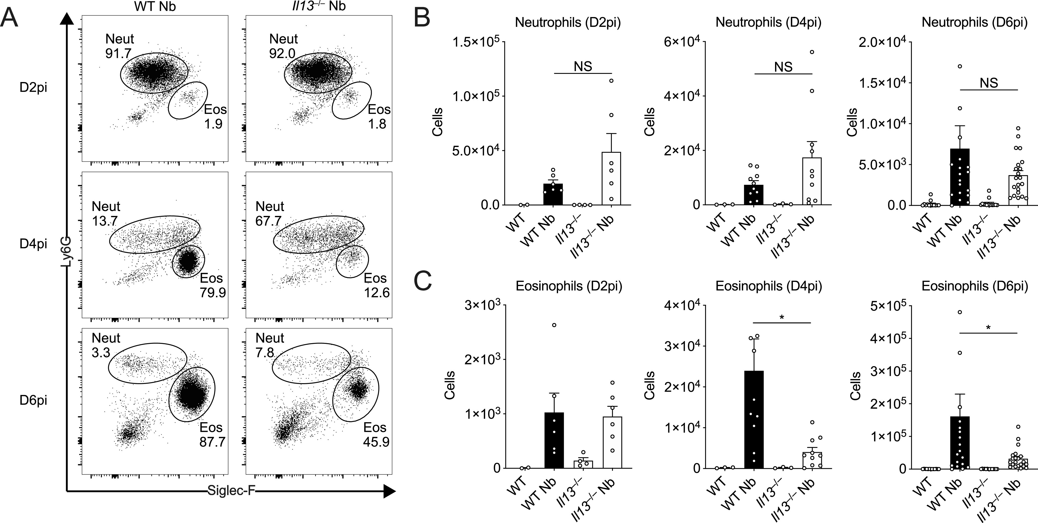
WT and Il13−/− mice were infected with N. brasiliensis (Nb) and BAL cells were analysed by flow cytometry. (A) Representative plots of percentages of BAL CD11c−CD11b+Ly6G+ neutrophils and CD11c−CD11b+Siglec-F+ eosinophils on D2, D4, and D6pi. Numbers indicate percentage of cells within total live CD45.2+ cells. (B, C) Total BAL neutrophil and (C) eosinophil cell counts on D2, D4, and D6pi. Data (mean ± SEM) were representative (day 2 post-infection) or pooled (D4 and D6pi) from four individual experiments with three to five mice per group (per experiment). NS: not significant, *P < 0.05 (one-way ANOVA and Tukey–Kramer post hoc test).