
Figure 3. IL-4 and IL-5 do not compensate in the absence of IL-13 during Nippostrongylus brasiliensis infection.
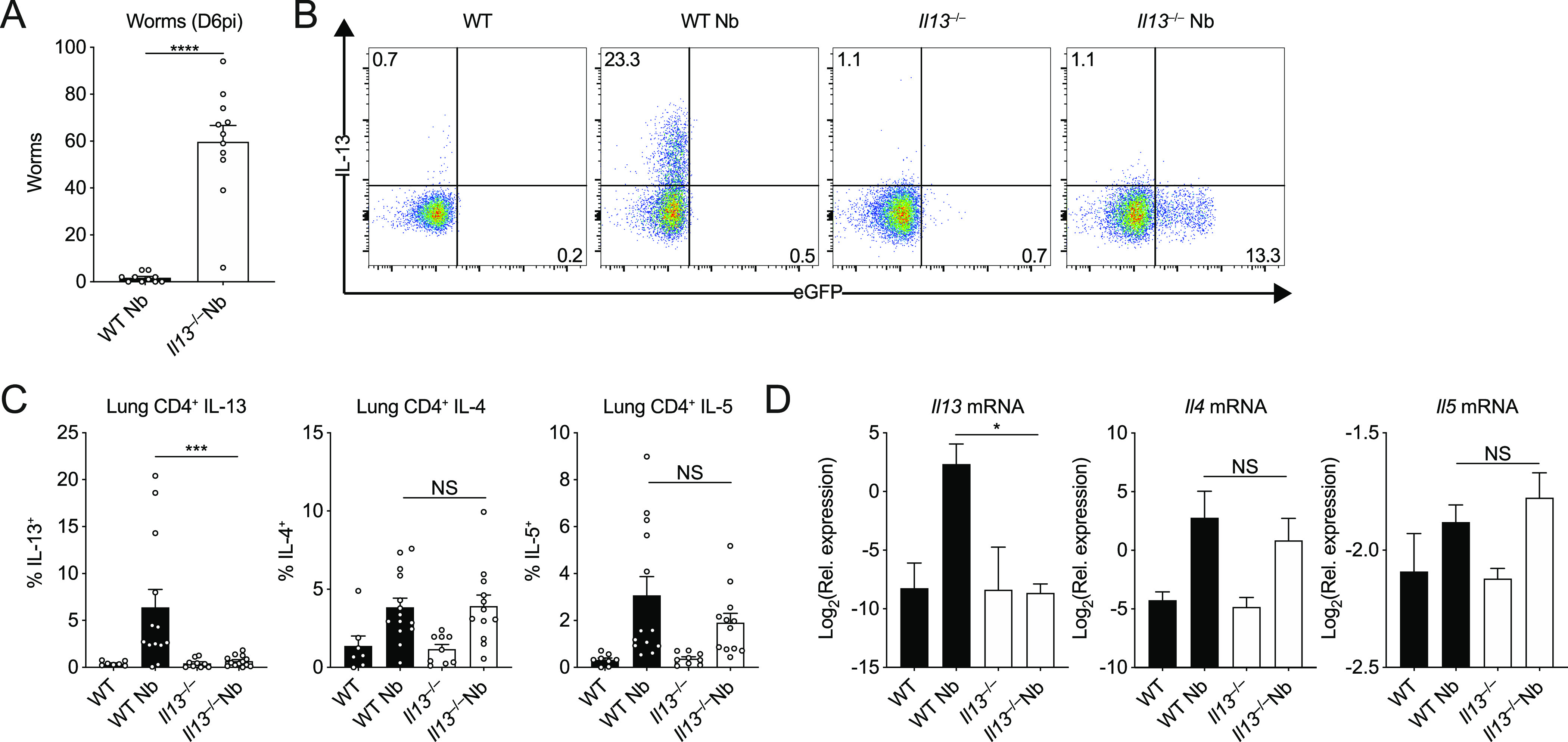
WT and Il13−/− mice were infected with N. brasiliensis (Nb). (A) On D6pi, adult worms in the small intestine were quantified. (B) Representative flow cytometry plots of lung CD4+ T cells stimulated ex vivo to measure intracellular WT IL-13 and KO eGFP expression. (C) Percentages of CD4+ T cells expressing IL-13, IL-4, and IL-5. (D) Whole lung Il13, Il4, and Il5 mRNA was measured by quantitative real-time PCR (data normalised against housekeeping gene Rpl13a). Data (mean ± SEM) were pooled from three individual experiments with three to five mice per group (per experiment). NS: not significant, *P < 0.05, ***P < 0.001, ****P < 0.0001 (one-way ANOVA and Tukey–Kramer post hoc test).