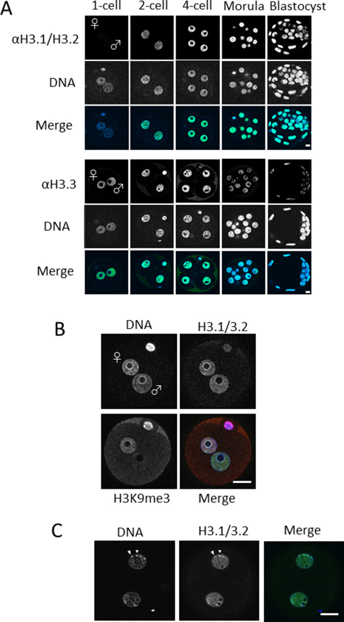
Figure 1. Nuclear deposition of histone H3 variants in mouse preimplantation embryos.
(A) One-cell, two-cell, four-cell, morula, and blastocyst-stage embryos were immunostained using anti-H3.1/2 (top half) and anti-H3.3 (bottom half) antibodies. Four to five independent experiments were performed. 8–15 embryos were observed for each developmental stage in each experiment; 39–64 embryos were analyzed in total. Representative images are shown for each experiment. Scale bar, 10 μm. (B) Enlarged images of stained one-cell embryos with enhanced confocal detector gain. In addition to H3.1/2, H3K9me3 was immunostained to discriminate the male and female pronucleus. In the merged panel, blue, green, and red colors represent the signals of DNA, H3.1/2, and H3K9me3, respectively. Scale bar, 20 μm. (C) Enlarged images of two-cell embryos; arrowheads indicate chromocenters. In the merged panel, blue and green colors represent the signals of DNA and H3.1/2, respectively. Scale bar, 20 μm.

