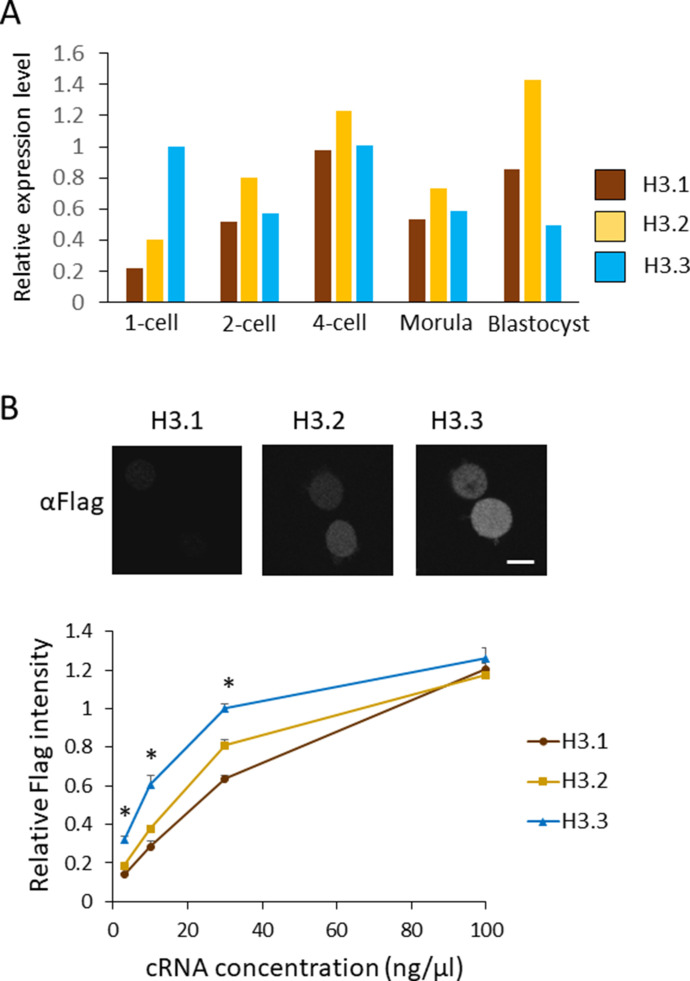
Figure 2. Nuclear localization of H3.1/2 is regulated by mRNA levels and histone incorporation efficiency in one-cell-stage embryos.
(A) mRNA expression levels of H3 variants during preimplantation development. RPKM values were obtained from previously published RNA-seq data (Abe et al, 2015). RPKM values for each gene encoding H3.1, H3.2, or H3.3 were totaled; the total RPKM of H3.3 at the one-cell stage was normalized to 1. (B) The incorporation efficiency of histone H3 variants into chromatin of one-cell embryos. Approximately 10 pl of H3.1, H3.2, or H3.3-FLAG cRNA was microinjected into MII-stage oocytes at various concentrations (3, 10, 30, and 100 ng/µl). After insemination, embryos were collected at 11 h post-insemination (hpi) and subjected to immunostaining. Anti-FLAG antibody was used to detect FLAG-tagged histones incorporated into chromatin. Representative immunocytochemistry images depict one-cell embryos, in which 10 ng/µl of H3.1, H3.2, or H3.3-FLAG was microinjected. Scale bar, 10 μm. The incorporation efficiency of H3 variants at one-cell embryos is shown as a line graph. The signal intensity for H3.3 microinjected at 30 ng/µl concentration was normalized to 1. Nine experiments were performed in total, using H3.3 injected with 30 ng/µl as a control for each experiment. Three to four experiments were performed for each concentration. Ninety 1-cell embryos were analyzed for the H3.3 30 ng/µl concentration. For embryos microinjected with other cRNA concentrations, 26–43 embryos were analyzed in total. Bars indicate standard error. Asterisks indicate that the value for H3.3 was significantly higher than both values of H3.1 and H3.2 (P < 0.01 by t test).

