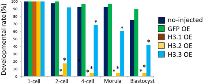
Figure S3. Impact of FLAG-tagged H3 variant overexpression on preimplantation development.
Mature oocytes were injected with 100 ng/µl H3.1, H3.2, or H3.3 with a FLAG-tag attached at the C terminus. Oocytes were fertilized; embryos with two pronuclei were determined to be one-cell embryos. Developmental progression was observed at the following time points: two-cell (28 h post-insemination [hpi]), four-cell (45 hpi), morula (72 hpi), and blastocyst (96 hpi). Three independent experiments were performed; 4–27 embryos were observed per group in each experiment, with a total of 29–46 embryos for each group. For H3.1- and H3.2-overexpressing embryos, a χ2 test or Fisher’s exact test (when there was a group in which the value was below 5) was performed; the results were considered significant when P < 0.01 for all noninjected, GFP-expressing, and H3.3-overexpressing embryos. For H3.3-overexpressing embryos, a χ2 test or Fisher’s exact test was performed; the results were considered significant when P < 0.01 for both noninjected and GFP-overexpressing embryos.

