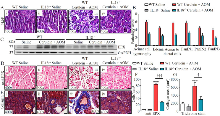
Figure 6. Improved malignant pathological phenotype detected in cerulein- and AOM-treated IL-18 gene–deficient mice.
(A) Representative photomicrographs show improved pancreatic pathology of acinar cell hypertrophy, accumulation of inflammatory cells, ductal hyperplasia, and formation of PanINs and periductal stroma in IL-18−/− mice compared with cerulein-with-AOM–treated murine model of chronic pancreatitis (Ai–iv). (B, C) Semi-quantitative pathology scores presented using light microscopic analysis (C). Highly reduced EPX protein expression by immunoblotting in IL-18−/− mice compared with cerulein-with-AOM–treated mice. (D, E) Immunohistology further confirmed reduced EPX positive cells in IL-18−/− mice compared to cerulein-with-AOM–treated murine model of chronic pancreatitis (E), Masson trichrome analysis for collagen staining showed reduced collagen area in IL-18−/− mice compared with cerulein-with-AOM–treated mice. (F, G) Morphometric analysis shows EPX+ cells and collagen area in IL18−/− mice with or without cerulein + AOM treatment. Data are expressed as means ± SD, n = 8 mice/group. All photomicrographs shown are in original magnification of 400× (scale bar 20 µm). †P < 0.05, *** or †††P < 0.0001. *Represents WT cerulein with AOM versus WT saline and AOM, † IL18−/− cerulein with AOM versus WT cerulein with AOM.

