Figure 4.
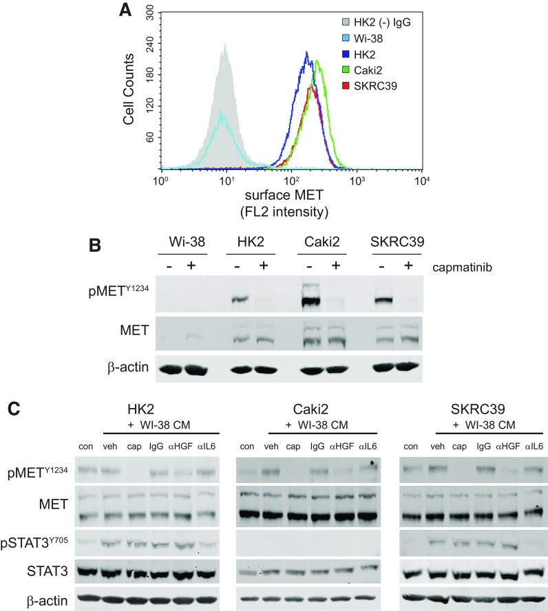
Fibroblast-derived hepatocyte growth factor (HGF) activates epithelial MET in papillary renal cell carcinoma (pRCC) cell tumor cells. A: flow cytometry histogram of MET receptor surface expression in Wi-38 fibroblasts, HK2 normal proximal tubule cells, and the two pRCC cell lines (Caki2 and SKRC39). HK2 cells stained with mouse isotype IgG were used to establish the background signal for the assay (gray shaded histogram). B: immunoblots of whole cell lysates for activated and total MET expression in all four cell lines indicated above. Actin was included as a control for protein loading. C: immunoblots of whole cell lysates after 30 min of treatment with the indicated conditions. Conditioned media (CM) from Wi-38 fibroblasts were harvested after 72 h of culture and then pretreated overnight with the indicated controls or inhibitors of HGF/MET signaling prior to treatment of renal cells. con, RPMI-1640 + 5% FBS; veh, 0.1% DMSO; CAP, 100 nM capmatinib; IgG, 1 µg/mL nonspecific mouse IgG; αHGF, 1 µg/mL human HGF neutralizing antibody; αIL6, 1 µg/mL interleukin-6 neutralizing antibody.

