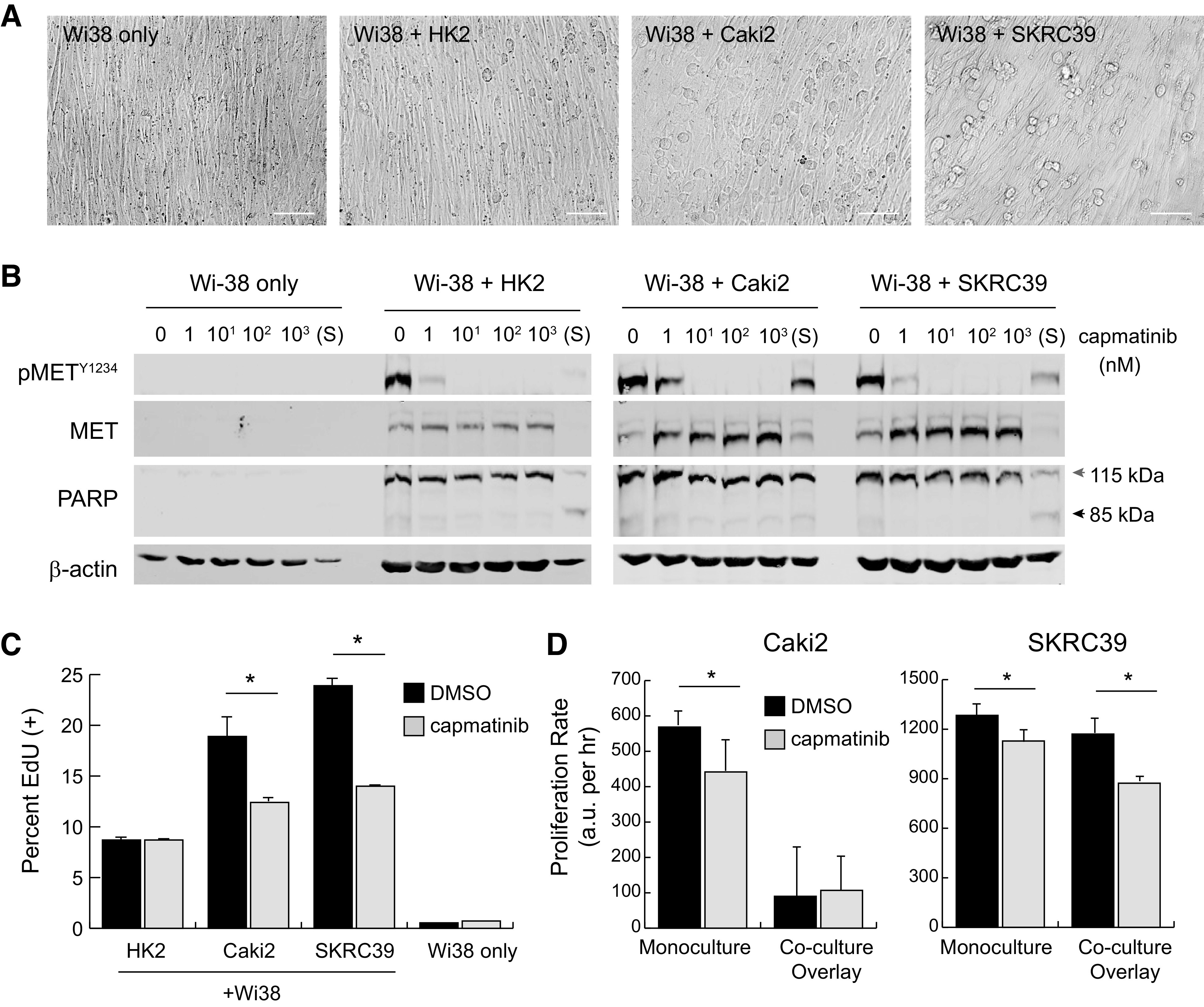
Figure 5.

Two-dimensional overlay coculture induces paracrine hepatocyte growth factor (HGF)/MET signaling but has limited impact on tumor cell proliferation. Wi-38 fibroblasts were grown to confluency in 24-well plates in serum-free media and then overlaid with 50,000 renal cells to create two-dimensional cocultures. A: images of different mono- or cocultures at 24 h postplating. Scale bar = 125 µm. B: immunoblots from whole cell lysates of two-dimensional cultures at 24 h postplating with the indicated nanomolar concentrations of capmatinib or after 1 h of treatment with 1 µM staurosporine (S). Poly(ADP-ribose) polymerase (PARP) cleavage from 115 to 85 kDa was included as an indicator of cell death; actin was included as a control for protein loading. C: active S-phase entry of coculture cells treated with capmatinib (100 nM) or vehicle (DMSO) at 72 h, as quantified by 5-ethynyl-deoxyuridine (EdU) incorporation. Values represent the average of three replicates with error bars indicating the standard deviation. Significance was determined by pairwise comparison for each set of samples (*P < 0.05). D: the proliferation rate of cocultures treated with capmatinib (100 nM, gray) or DMSO (black) was measured using a Cell Titer Glo luminescence assay. Luminescent values were captured from triplicate wells at regular intervals for 72−96 h and fit to linear equations to obtain growth rates with values of arbitrary luminescent units (a.u.) per hour. The significance of growth rate difference between vehicle and drug conditions is shown for each cell line (*P < 0.05).
