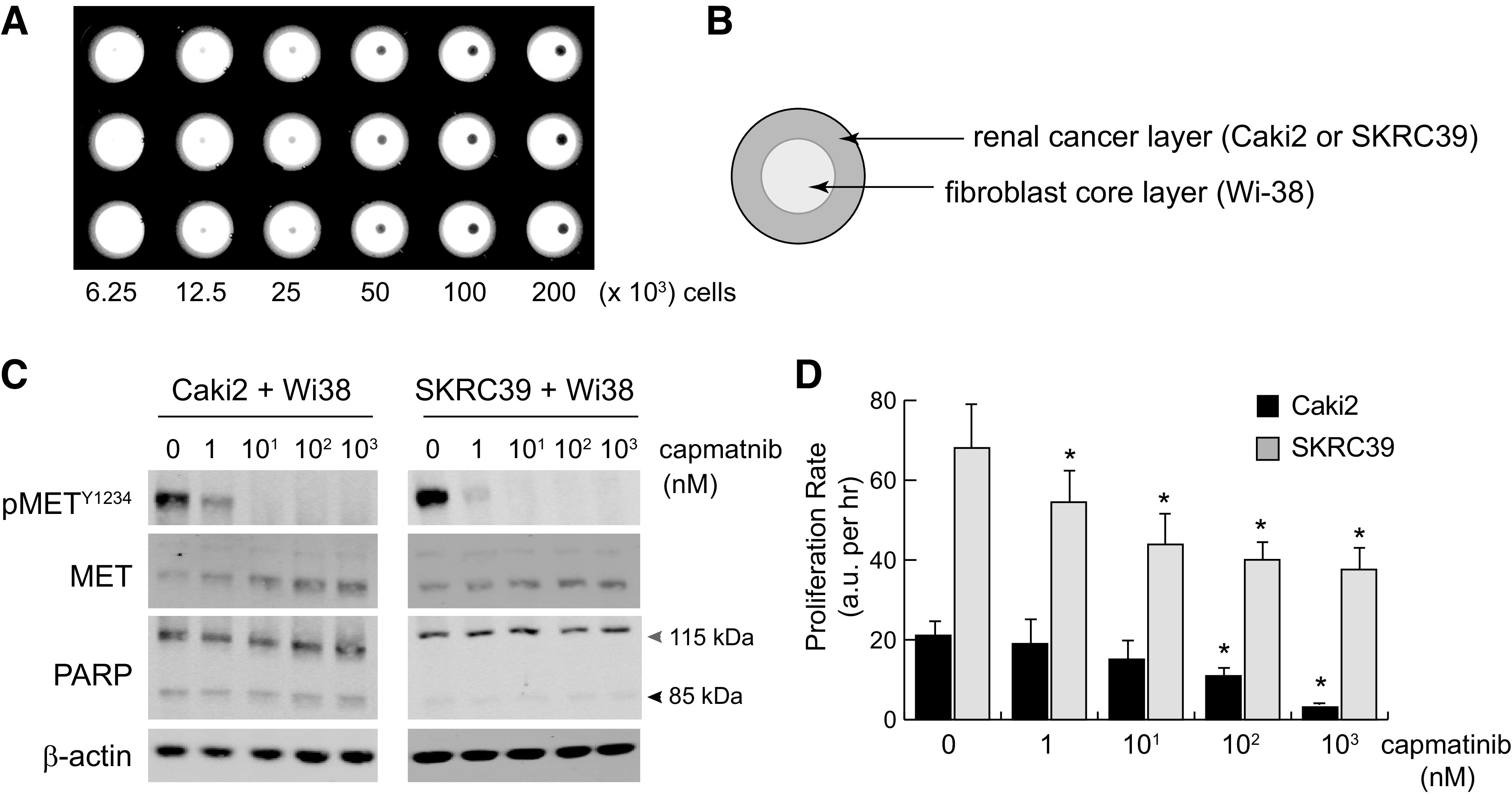
Figure 6.

Three-dimensional tumor spheroid coculture reproduces paracrine hepatocyte growth factor (HGF)/MET signaling and is sensitive to pharmacological MET inhibition. Three-dimensional spheroids were created by incubating magnetized cells in nonadherent 96-well plates placed above a magnet. Plates were removed from the magnet after 3 h to allow for normal spheroid compaction resulting from cell-cell adhesion. A: low-magnification image of spheroids plated at a twofold serial density of 200,000–6,250 cells/well. B: to create coculture spheroids, Wi-38 fibroblasts were used to form the inner core layer, which was then coated with an outer layer of cancer cells 3 h later. Cells were cultured in serum-free media with heparin to promote HGF production and paracrine signaling. C: immunoblots from single spheroid lysates at 24 h postplating with the indicated nanomolar concentrations of capmatinib. Poly(ADP-ribose) polymerase (PARP) cleavage from 115 to 85 kDa was included as an indicator of cell death; actin was included as a control for protein loading. D: the proliferation rate of cocultured spheroids treated with the indicated concentration of capmatinib was measured using a Cell Titer Glo three-dimensional luminescence assay. Luminescent values were captured from three individual spheroids for each condition at regular intervals for 72–96 h and fit to linear equations to obtain growth rates with values of arbitrary luminescent units (a.u.) per hour. The significance of growth rate difference for each condition was determined relative to the same line grown in vehicle only (0 nM) (*P < 0.05).
