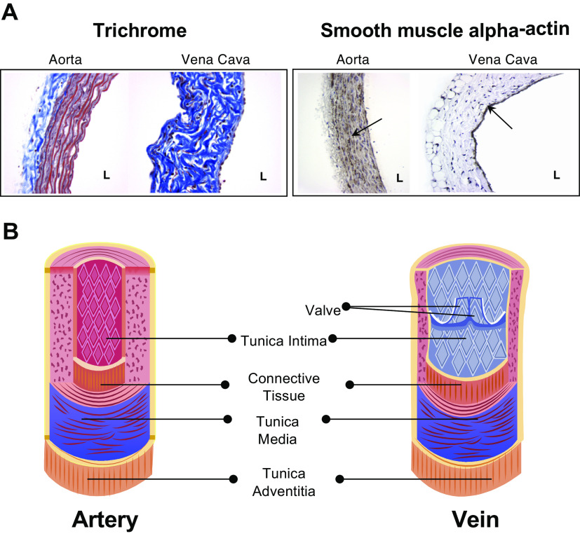
Figure 15.
A: three colors stain (Trichrome) and vascular smooth muscle cells (VSMC) alpha actin staining in the rat thoracic aorta vs. vena cava (above the liver). L, lumen. B: veins have valves to promote the one-way movement of blood from tissues back to the heart. Used with permission from Hartmannsgruber et al. (51).

