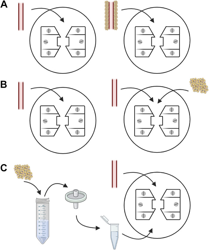
Figure 16.

Experimental protocols assessing the vasoactive effects of perivascular adipose tissue (PVAT). Two segments of the same vessel are tested, with one segment being PVAT-denuded and the other PVAT-intact (A). A vascular segment denuded from PVAT is mounted into a wire myograph and is tested in the absence of PVAT (left). Subsequently, the same vascular segment is tested in the presence of its neighboring PVAT (right), which had been initially removed and stored at 4°C physiological salt solution until experimentation. In this protocol, PVAT from comparing groups and conditions can be used to determine whether the observed effect is specific to (patho)physiological state (B). A vascular segment is cleaned from its PVAT, which is incubated in physiological salt solution at 37°C to create PVAT-conditioned media. The PVAT solution is filtered and added into the wire myograph chamber. Concentration-response curves to selected stimuli are performed in the absence and presence of PVAT-conditioned media (C). Created with Biorender.com.
