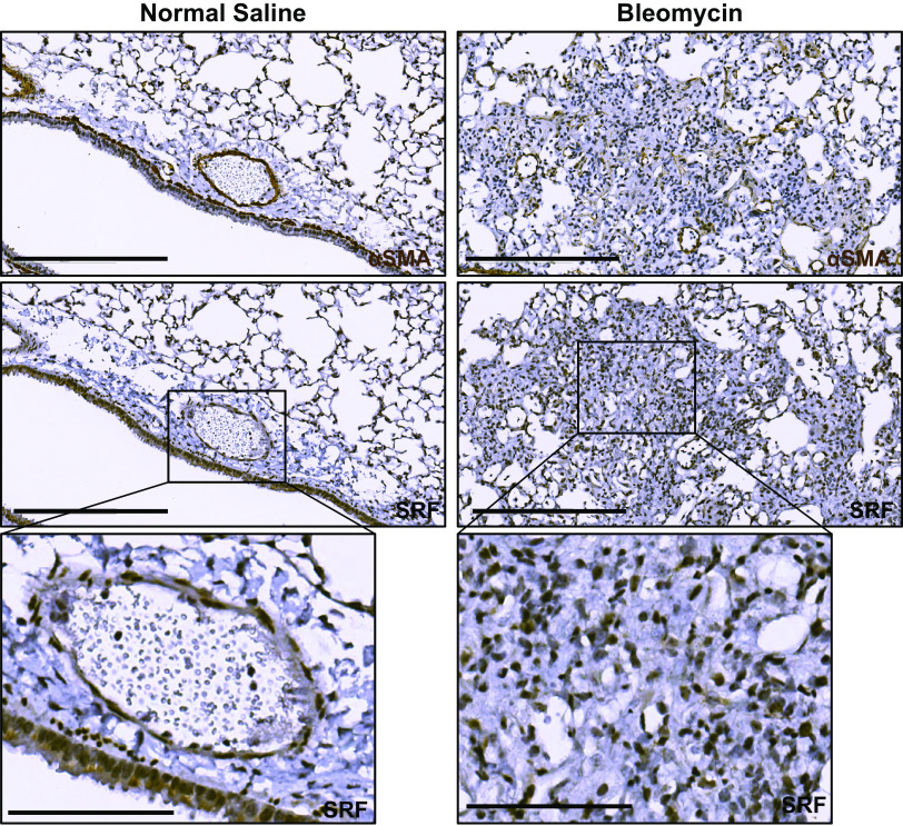
Figure 2.
Serum response factor (SRF) expression is increased in the fibrotic regions of bleomycin-induced pulmonary fibrosis. Wild-type mice were intratracheally treated with 0.9% normal saline (left column) or 1 U/kg bleomycin (right column) (n = 3 animals for each condition). Fourteen days later, mice were euthanized and isolated lungs were fixed with 10% neutral buffered formalin, paraffin embedded, and subject to immunohistochemical staining against smooth muscle α actin (αSMA) and SRF. Left column: normal saline-treated, uninjured mouse lungs stained against αSMA (first row) showed increased expression surrounding airways and blood vessels. Matching histologic areas demonstrate increased SRF staining in the smooth muscle layer of blood vessels and airways (matching αSMA expressing cells), as well as faint staining in the columnar epithelium of the airway (row two and three). In fibrotic mouse lung (right column), there is increased αSMA staining of consolidated and fibrotic lung parenchyma (first row). SRF immunostaining of matched histologic samples shows increased staining in the same areas of the fibrotic parenchyma (second and third rows). Harris hematoxylin counterstain was performed for nuclear labeling in all stains. Scale bar = 300 μm (rows 1 and 2). Third row: digitally zoomed regions of increased expression of SRF in the blood vessel and part of airway (left, normal) and fibroblastic focus (right, fibrotic). Scale bar = 100 μm (row 3).

