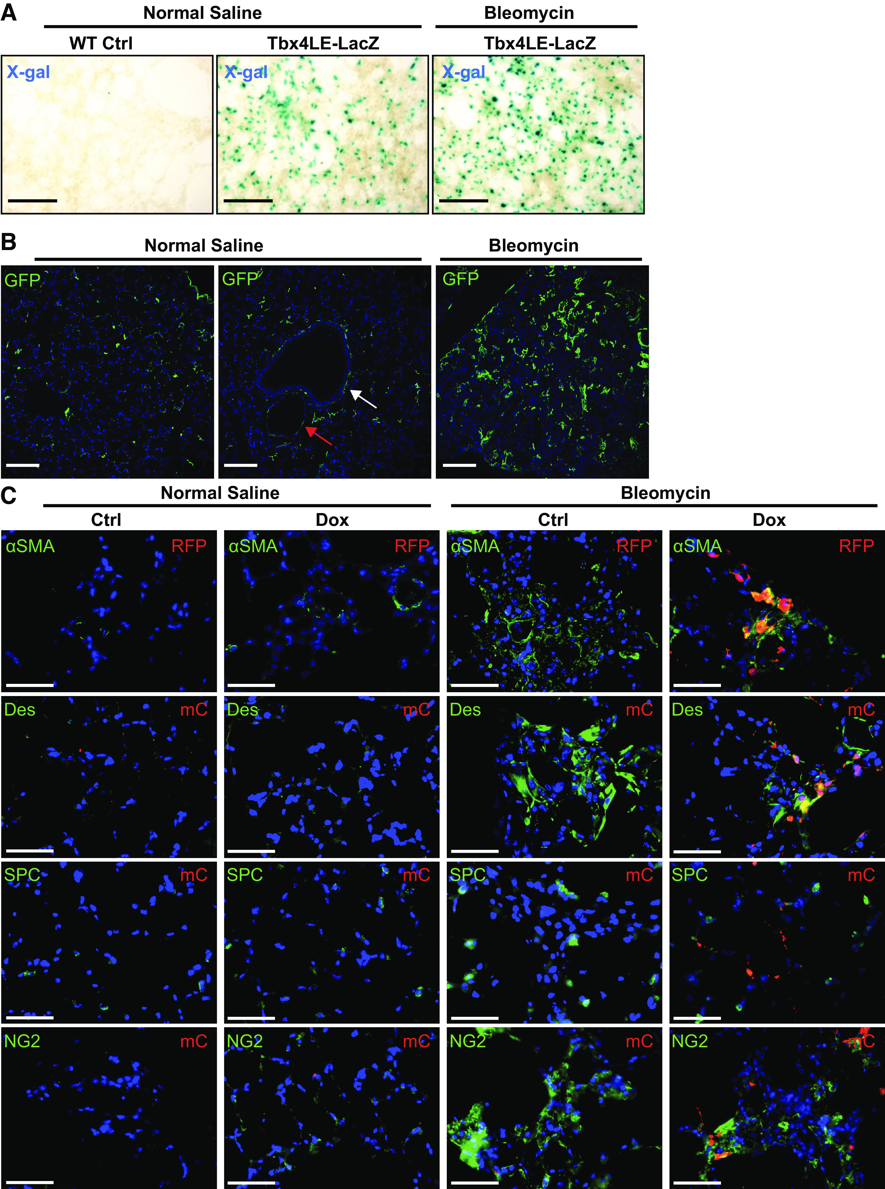
Figure 3.

Lung mesenchymal cells expand and express myofibroblast markers postbleomycin treatment. A: Tbx4LE-rtTA-LacZ-expressing mice or wild-type (WT) controls were subject to intratracheal bleomycin (1 U/kg suspended in 0.9% normal saline, NS) or NS treatment. Eight days after bleomycin treatment, 150-μm-thick precision cut lung slices were stained with X-gal and imaged. Representative images are displayed for each condition. B: Tbx4LE-rtTA/TetO-Cre/mTmG mice were treated with doxycycline (2 mg/mL + 50 mg/mL sucrose in water), and 2 days later, subject to intratracheal bleomycin or NS treatment. Fourteen days after bleomycin instillation, lungs were fixed with 10% neutral buffered formalin, paraffin embedded, and stained against green fluorescent protein (GFP). Red arrow points to a blood vessel and white arrow points to an airway (middle). C: Tbx4LE-rtTA/TetO-Cre/tdTom mice were given doxycycline (dox) in water (2 mg/mL + 50 mg/mL sucrose) or sham (50 mg/mL sucrose) as a control starting 2 days before a single intratracheal bleomycin or NS treatment. Fourteen days after bleomycin instillation, mouse lungs were fixed with 10% neutral buffered formalin, paraffin embedded, and subject to immunofluorescence double staining against red fluorescent protein (RFP) or mCherry (mC) to enhance tdTomato signal and smooth muscle α actin (αSMA), Desmin (Des), prosurfactant protein C (SPC), or neural/glial antigen 2 chondroitin sulfate proteoglycan (NG2). All fluorescence images are counterstained with 4′,6-diamidino-2-phenylindole (DAPI) (blue). In all images, scale bar = 100 µm.
