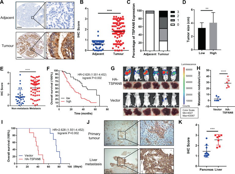
Fig. 1. TSPAN8 is highly expressed in PDAC and is associated with progression and poor prognosis.
A–C Immunohistochemical staining (A), TSPAN8 expression (B–C) and tumor size (D) data for 87 human pancreatic cancer specimens were analyzed. Representative images of normal adjacent tissues (NATs) and tumor tissues (TTs) are shown. Scale bars: 200 μm. E TSPAN8 expression in tumor tissues from patients with distant metastasis and without distant metastasis was analyzed. F The survival times of 87 PDAC patients with low (black curve) and high (red curve) TSPAN8 protein levels (low, 47 patients; high, 40 patients) indicated a significant association of the TSPAN8 level with patient survival, as determined by a log-rank test. G–H A total of 106 SW1990 cells with or without expression of HA-TSPAN8 were injected into athymic nude mice. Representative tumor xenografts are shown (G). The number of visible metastatic lesions in the liver was analyzed by a t test (H). I Kaplan–Meier survival analysis was performed. P values were calculated by a log-rank test (N = 6 mice). J–K Immunohistochemical staining for TSPAN8 was performed on 17 PDAC patient specimens of primary tumor tissues and liver metastases (J). Scale bars: 200 μm. TSPAN8 expression in primary tumor tissues and liver metastasis tissues was analyzed by a t test (K). B, D, E, H, K the values are presented as the means ± SDs. *P < 0.05, **P < 0.01, ***P < 0.001 and ****P < 0.0001.

