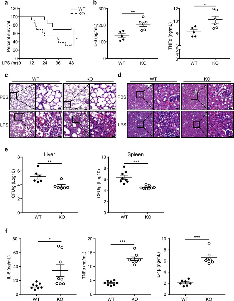
Fig. 6. TRIM60 deficiency potentiates the TLR-mediated innate immune response in vivo.
a Age-matched WT (n = 13) and Trim60−/− (n = 13) mice were intraperitoneally injected (i.p.) with LPS (50 mg/kg body weight), and the survival was monitored for 48 h, followed by Kaplan–Meier analysis. b ELISA analysis of the IL-6 and TNFα levels in sera from WT (n = 5) and Trim60−/− mice (n = 6) 6 h after the LPS injection. Each dot represents an individual mouse treated as indicated. H&E staining of lung (c) and kidney (d) tissues from WT and Trim60−/− mice injected with LPS. e, f Six-week-old WT (n = 8) and Trim60−/− (n = 8) mice were infected via the i.v. route with L. monocytogenes at 2 × 10⁴ CFU. The bacterial loads in the liver and spleen were examined at 72 h after infection (e). The proinflammatory cytokine levels in sera were tested at 24 h after infection (f). Each dot represents an individual mouse infected with L. monocytogenes. The data in b, e, and f are presented as the mean ± SEM. *P < 0.05; **P < 0.01; ***P < 0.001 (log-rank test for a; two-tailed unpaired Student’s t test for b, e, f). The scale bars in c and d) represent 20 μm. The results in c and d are representative of four pairs of WT and TRIM60 KO mice

