Fig. 3.
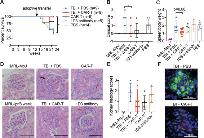
CAR-T cell treatment shows therapeutic benefits in mouse SLE. Mice received different treatments at 13 weeks of age and were sacrificed at 22 weeks of age for subsequent examinations. a The survival rate of MRL-lpr mice that received different treatments; the difference in survival between the TBI + PBS group and TBI + CAR-T group was compared by logrank (Mantel–Cox test) calculation. b The mouse skin lesions were scored, and the scores were plotted. c The ratios of spleen weight to total body weight were determined. d Representative hematoxylin and eosin (H&E)-stained sections of glomerular areas of the kidneys. Black arrow, immune complex formation; red arrow, infiltration of numerous lymphocytes. Original magnification, ×400. Bars represent 50 µm. e Statistical histology scores of H&E-stained kidney sections. f C3 and IgG deposition in the glomerulus was detected by immunofluorescence and is presented in a merged manner; frozen sections of the kidneys were stained with anti-mouse-C3-APC, anti-mouse-IgG-FITC, and DAPI. Original magnification, ×400. Bars represent 50 µm. The data are representative of at least two independent experiments with similar results. b, c The data were analyzed by Student’s t test, and significance is indicated by *P < 0.05

