Fig. 4.
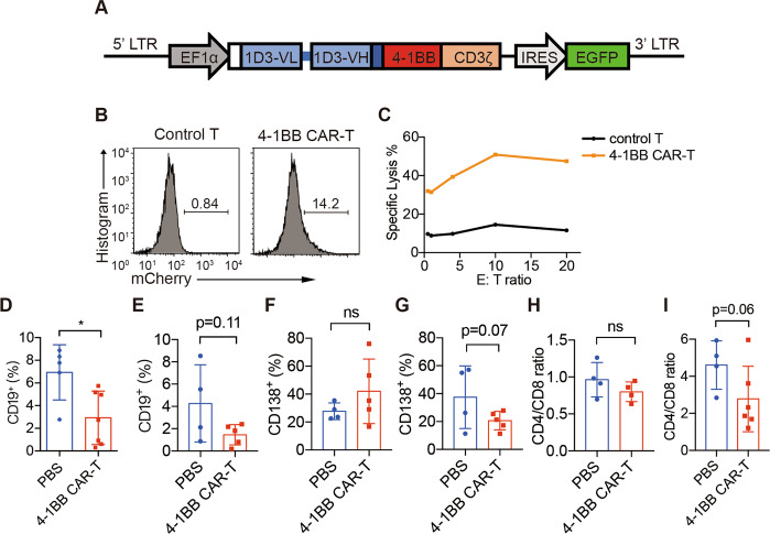
1D3-4-1BB CAR-T cells partially deplete B cells. a Diagram of the DNA encoding 1D3-4-1BB CAR. It encodes the antibody light chains (blue boxes), including their native signal peptides (white box), joined by a flexible linker (blue bar) to the VH and fused to the transmembrane region of CD8a (navy blue), signaling domains of 4-1BB (red box) and cytosolic domain of CD3ζ (orange box). The construct also contains IRES-driven EGFP to allow the detection of transduced cells. b Flow cytometry of GFP+ expression on mouse primary T cells after lentiviral transduction. Cells were FVD− TCRβ+ gated. c Unsorted 4-1BB CAR-T cells (effector) and CellTrace Violet-labeled splenocytes (target) were cocultured for 4 h and analyzed by flow cytometry. Blood was collected from the femoral arteries of MRL-lpr mice at 1 week (d, f) or 9 weeks (e, g) after transfer, and CD19+ and CD138+ percentages among FVD− CD45+ cells were analyzed by flow cytometry. CD4/CD8 ratios in the blood were also analyzed by flow cytometry at 1 week (h) or 9 weeks (i) after transfer. The data are representative of at least two independent experiments with similar results. The data were analyzed by Student’s t test, and significance is indicated by *P < 0.05

