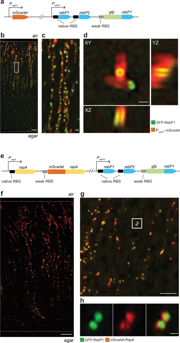
Fig. 4. RebP1 forms internal rings and co-localizes with RapA.
a Schematic of constructs present in the strain engineered to produce GFP-tagged RebP1, showing the PrebP1-mScarlet transcriptional reporter and the gfp-rebP1 translational fusion. b Representative confocal image of a thin section prepared from a 3-day-old biofilm formed by the strain indicated. Dotted lines demarcate the edges of the biofilm. Scale bar is 10 µm. c Higher-magnification image of the boxed region shows R-bodies within each cell. Scale bar is 1 µm. d Zooming in on single cells reveals fluorescently tagged R-bodies forming a characteristic ring structure. Orthogonal projections reveal the tubular open organization of GFP-RebP1. Scale bar is 500 nm. Note that the non-isotropic resolution slightly deforms the structure along the z axis. e Schematic of constructs present in the strain engineered to produce GFP-tagged RebP1 and mScarlet-tagged RapA, showing the gfp-rebP1 translational fusion and the mScarlet-rapA translational fusion. f Representative confocal image of a thin section prepared from a 3-day-old biofilm formed by the strain indicated. Dotted lines demarcate the edges of the biofilm. Scale bar is 10 µm. g Super-resolution imaging reveals colocalization of mScarlet-RapA with the R-body ring structure formed by GFP-RebP1. Scale bar is 5 µm. h Zoom-in of the boxed region shown above. Scale bar is 500 nm. Images are representative of three fields of view from two experiments.

