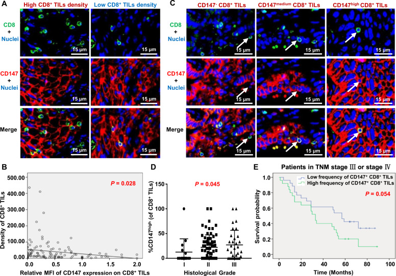
Fig. 8.
Correlations between the CD147 expression on CD8+ TILs and clinicopathological characteristics of non-small-cell lung cancer (NSCLC) patients. A Representative figures of double-immunofluorescence staining for CD147+ CD8+ TILs in NSCLC tumor tissue samples with a high or low CD8+ TIL density using anti-CD8a (green) and anti-CD147 (red) antibodies. The nuclei were stained with DAPI (blue). B The correlation between the MFI of CD147 staining on CD8+ TILs and the CD8+ TIL density was determined with linear- regression analysis (n = 117). C Representative images of CD147− CD8+, CD147medium CD8+, and CD147high CD8+ TILs. Arrow, a representative CD8+ TIL. D The correlation between the frequency of intratumoral CD147high CD8+ TILs and histological grade was determined with Spearman correlation analysis (n = 117). E The overall survival of NSCLC patients with TNM-stage III or IV disease was compared between the group with a high frequency of CD147+ CD8+ TILs (>24.64%) and the group with a low frequency (≤24.64%) with the Kaplan–Meier method and log-rank test (n = 51)

