Figure 1.
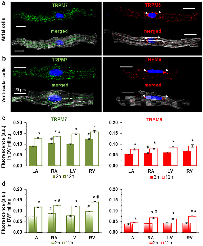
Immunofluorescence of TRPM7 and TRPM6 proteins in all cells used. Image acquisition performed using confocal laser scanning microscope (a, atria; b, ventricle). Immunofluorescence of confocal z-stack of cardiomyocytes with immunodetected TRPM7 and TRPM6 proteins, respectively. Alexa Fluor 488 and Alexa Fluor 546 for the TRPM7 and TRPM6 protein appear in green and red, respectively. Alexa Fluor 405 for F-actin cytoskeleton appears in surrogate grey. Hoechst 33342 for nuclei appears in blue (the arrowheads indicate the localization of TRPM6 protein in the perinuclear area). (c, d) Quantification of immunofluorescence levels of the TRPM7 (green) and TRPM6 (red) proteins in cardiomyocytes from four chambers of the heart (left atrium, LA; right atrium, RA; left ventricle, LV; and right ventricle, RV), under experimental conditions with (c) and without (d) divalent cations in the extracellular milieu, respectively. Cardiomyocytes were fixed following 2 h (filled columns) or 12 h (unfilled columns) after cell isolation. Mean data provided in arbitrary units (a.u.) (Supplementary Table 1 online). A blinded study-design (with the investigator reading the fluorescence not knowing the cell incubation conditions) was used for the detection of protein concentration during various experimental conditions. *P < 0.05 2 h vs. 12 h and #P < 0.05 right-sided vs. left-sided heart chambers. Scale bars indicate 20 µm.

