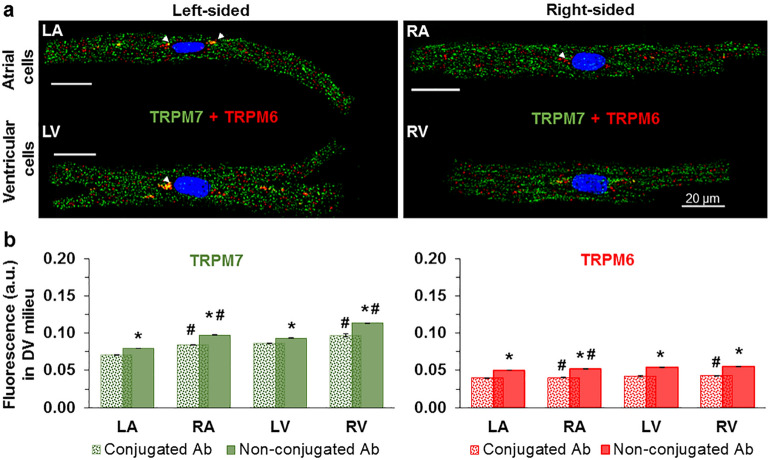
Figure 2.
Immunofluorescence images depicting co-expression of TRPM6 and TRPM7 proteins in human cardiomyocytes. (a) The immunofluorescence of TRPM7 (green) and TRPM6 (red) in the same LA, RA, LV, and RV cardiomyocyte when using conjugated antibodies (the arrowheads indicate the localization of TRPM6 protein in perinuclear area). (b) Quantification of the staining intensity of the immunodetected conjugated antibodies (spotty) and non-conjugated antibodies (smooth) for both proteins in cardiomyocytes from the four chambers of the heart as indicated. *P < 0.001 2 h vs. 12 h, #P < 0.001 right-sided vs. left-sided heart chambers. Other notations are the same as in Fig. 1.

