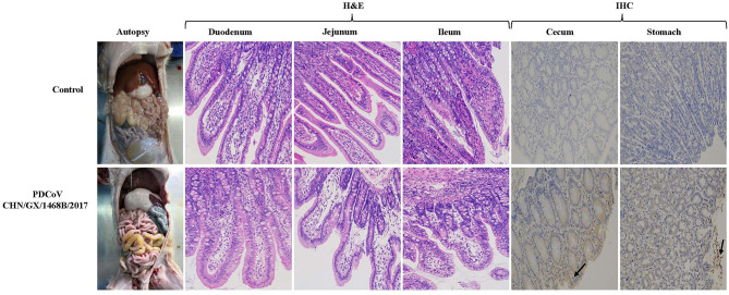
Figure 4.
Gross pathological, histopathological and immunohistochemical staining in piglets infected with the PDCoV CHN/GX/1468B/2017 strain. Typical macroscopic intestinal lesions were observed in piglets infected with the PDCoV CHN/GX/1468B/2017 strain by necropsy examination. The small intestine was expanded, filled with yellow water-like liquid and the intestinal wall became thin and transparent. H&E staining showed severe intestinal damage in infected piglets, especially in the small intestine. Obvious visible lesions were observed in the jejunum and ileum, and the villi from these regions were severely atrophied and blunted. In addition, the superficial villi on the epithelial cells were swollen and vacuolated. The duodenal cells were swollen and vacuolated. PDCoV antigen was detected by IHC using PDCoV-N protein specific polyclonal antibody and HRP-labeled goat anti-mouse IgG antibody. PDCoV antigen signals with brown coloring were detected in the cecum and stomach of piglets infected with the CHN/GX/1468B/2017 strain (indicated with black arrows). No antigen was detected in control piglets. (200× magnification).

