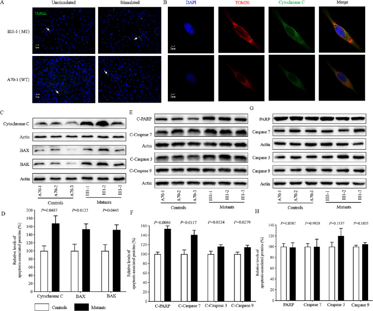
Figure 7.
Apoptosis assays. (A) TUNEL assays of mutant III1-1 and control A70-1 cybrids with or without H2O2 stimulation. Arrows indicate death cells. (B) The distribution of cytochrome c from mutant III1-1 and control A70-1 cybrids was visualized by immunofluorescent labeling with TOM20 antibody conjugated to Alexa Fluor 594 (red) and cytochrome c antibody conjugated to Alexa Fluor 488 (green) and analyzed by confocal microscopy. DAPI-stained nuclei were identified by their blue fluorescence. (C, E, G) Western blotting analysis. Cellular proteins (20 µg) from various cell lines were electrophoresed, electroblotted, and hybridized with several apoptosis-associated protein antibodies: cytochrome c, BAK, and BAX (C); cleaved caspases 3, 7, and 9 and PARP (E); and uncleaved caspases 3, 7, and 9 and PARP (G), with β-actin as a loading control. (D, F, H) Quantification of apoptosis-associated proteins: cytochrome c, BAK, and BAX (D); cleaved caspases 3, 7, and 9 and PARP (F); and uncleaved caspases 3, 7, and 9 and PARP (H). The levels of apoptosis-associated proteins in various cell lines were determined as described elsewhere.11 Three independent determinations were made in each cell line. Graph details and symbols are explained in Figure 1.

