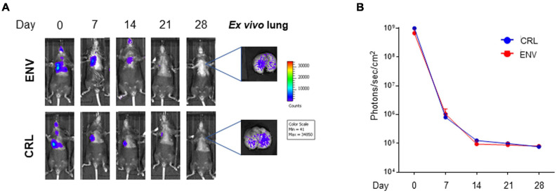
FIGURE 4.
In vivo and ex vivo imaging of intratracheally (IT) injected mouse bone marrow-derived mesenchymal stromal/stem cells (mBM-MSCs) from different sources. (A) In vivo bioluminescence imaging (BLI) of IT mBM-MSC clone #2LucRi/GFP inoculated Charles River Laboratories (CRL) and Envigo (ENV) mice. Mice were weekly monitored up to 4 weeks post inoculation. On day 28, mice were anesthetized and lungs were harvested for ex vivo imaging using In Vivo Imaging System (IVIS) BLI system. No significant differences were detected between the two strains of mice, thus host genetic background does not impact cell persistency into the lung (p > 0.05, two-way ANOVA test followed by Sidak’s test for multiple comparisons). (B) BLI signal from each mouse was quantified five times, expressed as photons/s/cm2 and plotted as mean ± SEM. BLI signal at each time point represents the mean ± standard deviation of five animals.

