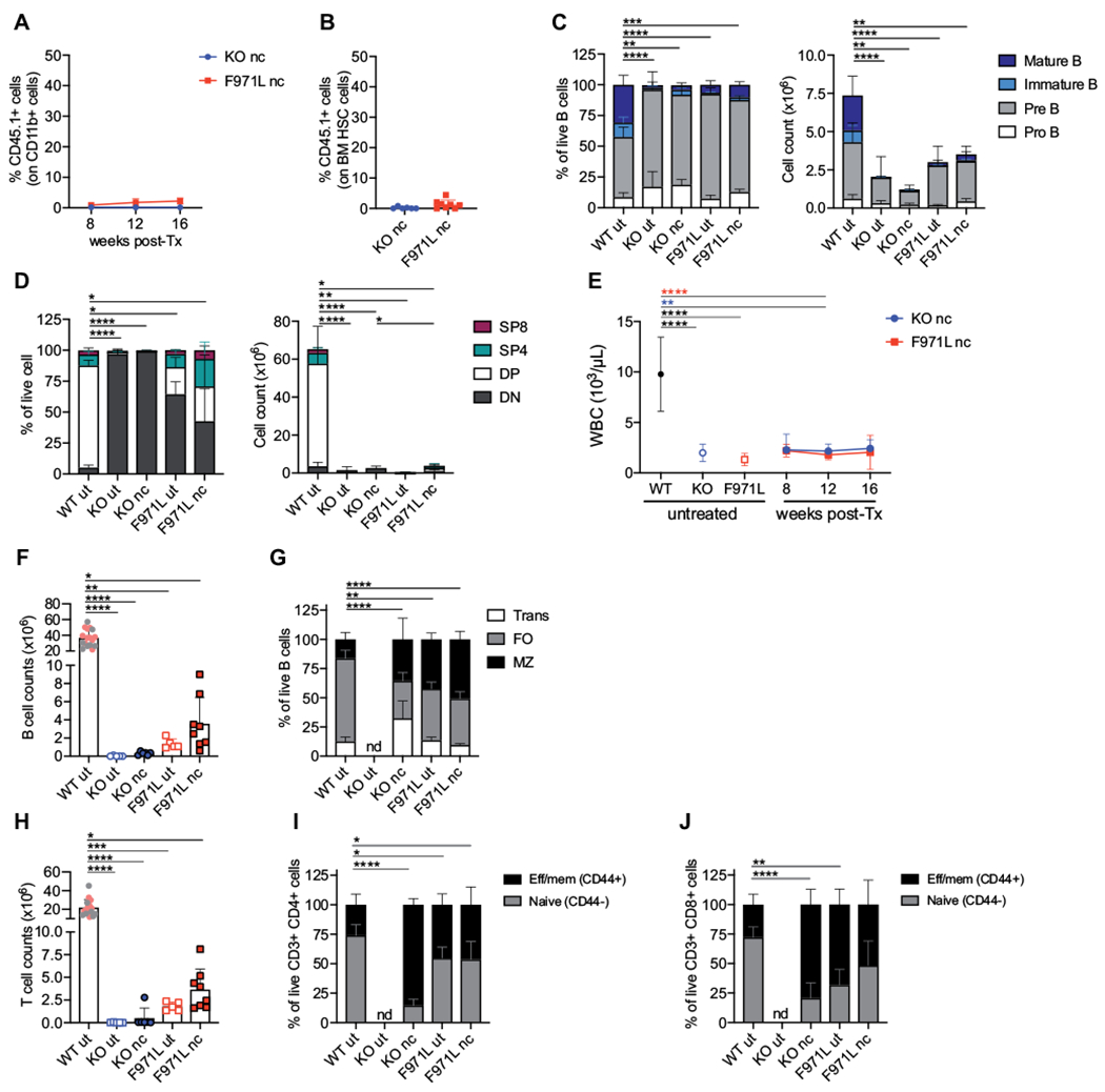
FIG 1.

Myeloid chimerism and immunologic reconstitution in unconditioned Rag1mut mice. A, Kinetics of myeloid chimerism measured by flow cytometry as the proportion of donor WT CD45.1+ cells gated on CD11b+ cells in the peripheral blood at different time points after the transplant (post-Tx) in nonconditioned (nc) Rag1-KO (knockout [KO]) mice (n = 6) and Rag1-F971L (F971L) mice (n = 5-9). B, Donor chimerism in HSCs identified as LSK CD150+CD48− cells of Lin− cells isolated from BM cells 16 weeks after the transplant. C, B-cell developmental stages were analyzed by flow cytometry in untreated (ut) mice or 16 weeks after transplant in nc mice and shown as proportion (left panel) and absolute counts (right panel) of B cells in BM (WT [n = 23], KO ut [n = 9], KO nc [n = 6], F971L ut [n = 5], and F971L nc [n = 8] mice). One-way ANOVA, Kruskal-Wallis test on pre–B-cell frequencies (left panel) and immature B-cell counts (right panel). D, CD4−CD8− double-negative (DN), CD4+CD8+ double-positive, SP4 (CD4+CD8−), and SP8 (CD4−CD8+) cells were analyzed by flow cytometry and shown as proportion of thymocytes (left panel) and absolute counts (right panel) (WT [n = 20], KO ut [n = 7], KO nc [n = 6], F971L ut [n = 5], and F971L nc [n = 8] mice). One-way ANOVA, Kruskal-Wallis test on DN cell frequencies (left panel) and double-positive (DP) cell counts (right panel). E, WBCs were measured over time in the PB. One-way ANOVA, Kruskal-Wallis test at 12 weeks after the transplant. Asterisk colors indicate the groups of comparison. F, Splenic B-cell counts are shown in the graph. G. Proportions of splenic transitional (Trans), follicular (FO), and MZ B cells were analyzed by flow cytometry. One-way ANOVA, Kruskal-Wallis test on FO B-cell frequencies. H, Splenic T-cell counts are shown in the graph. I and J, Phenotypic analysis results of splenic T lymphocytes are shown in the graph according to the expression of CD44 on the CD3+CD4+ cells (I) and CD3+CD8+ cells (J); 1-way ANOVA, Kruskal-Wallis test on naive T cells. F and H, Gray dots in WT bar were values from the SR-Tiget laboratory, and pink dots indicate values from the National Institutes of Health laboratory; 1-way ANOVA, Kruskal-Wallis test for multiple comparison. *P < .05; **P < .005; ***P < .0005; ****P < .0001. Means ± SDs are shown. Eff/mem, Effector/memory.
