
Extended Data Figure 4. MCU deficiency does not affect canonical autophagy.
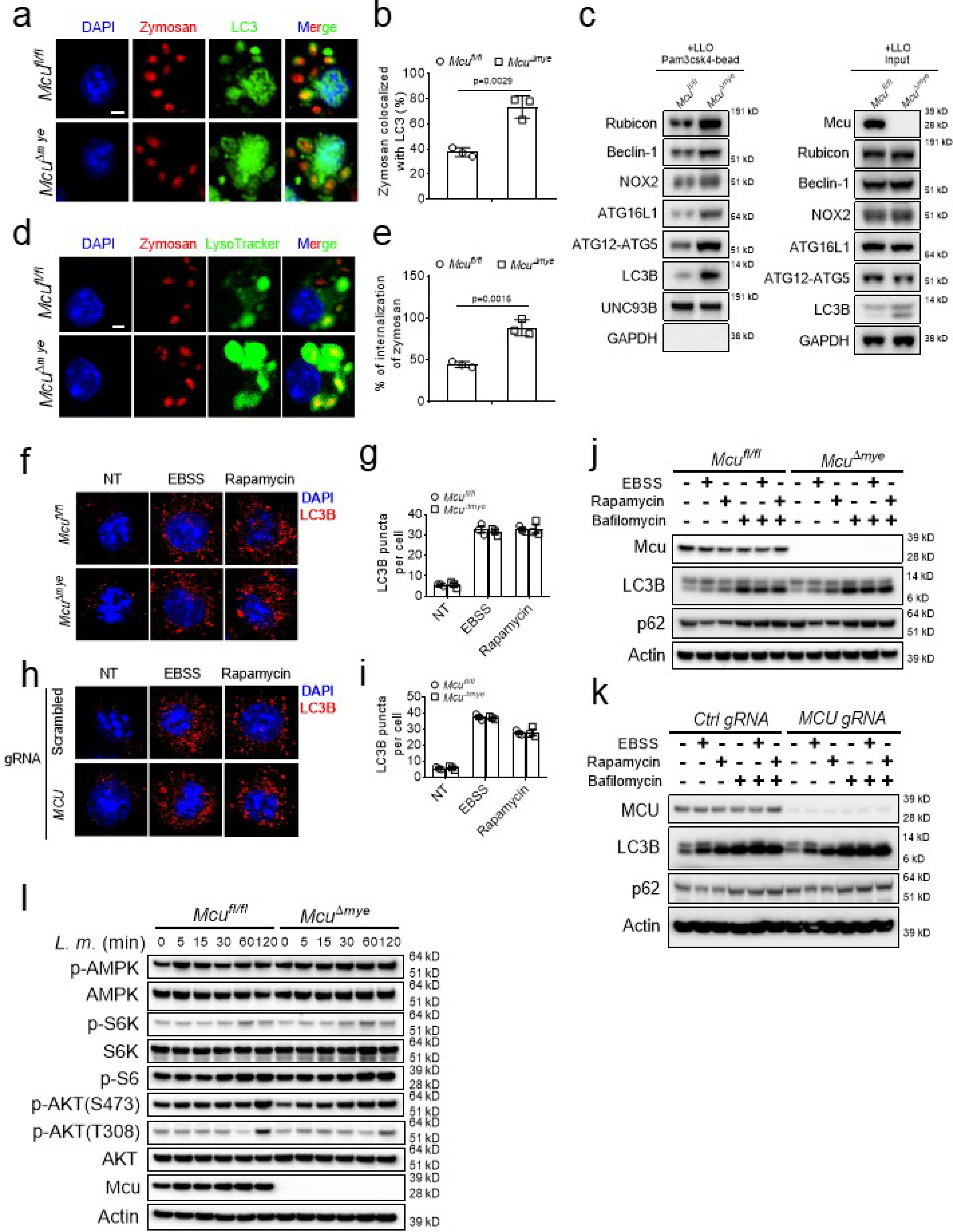
(a-e) Confocal imaging (a and d) and quantification (b and e) of the colocalization of Zymosan (red) with either LC3B puncta (green) (a and b) or LysoTracker (green) (de), immunoblotting of LAP-associated molecules in isolated phagosomes or total cell lysates from Mcufl/fl and McuΔmye BMMs in the presence of LLO (5 nM) (c). Scale bar, 2 μm. (f-i) Immunofluorescence staining of LC3B in Mcufl/fl and McuΔmye BMMs (f and g), or THP-1 MCU-WT and MCU-KO cells (h and i) left untreated or challenged. with EBSS for 2 h, or rapamycin (100 nM) for 16 h. LC3B puncta per cell (g and i) were shown. Scale bar, 2 μm. (j-l) Immunoblotting of LC3B and p62 in Mcufl/fl and McuΔmye BMMs (j), or THP-1 MCU-WT and MCU-KO cells (k) left untreated or pretreated with bafilomycin (50 nM) for 1 h, followed by EBSS or rapamycin (100 nM) incubation for another 2 or 16 h, respectively. Immunoblotting of AMPK and mTOR signaling molecules AKT, S6K and S6 in Mcufl/fl and McuΔmye BMMs challenged with L. monocytogenes (MOI, 10) for indicated periods (l). The averages of n = 3 biologically independent samples are shown. The error bars represent the SEM. Statistical significance was determined using t test (and nonparametric tests).