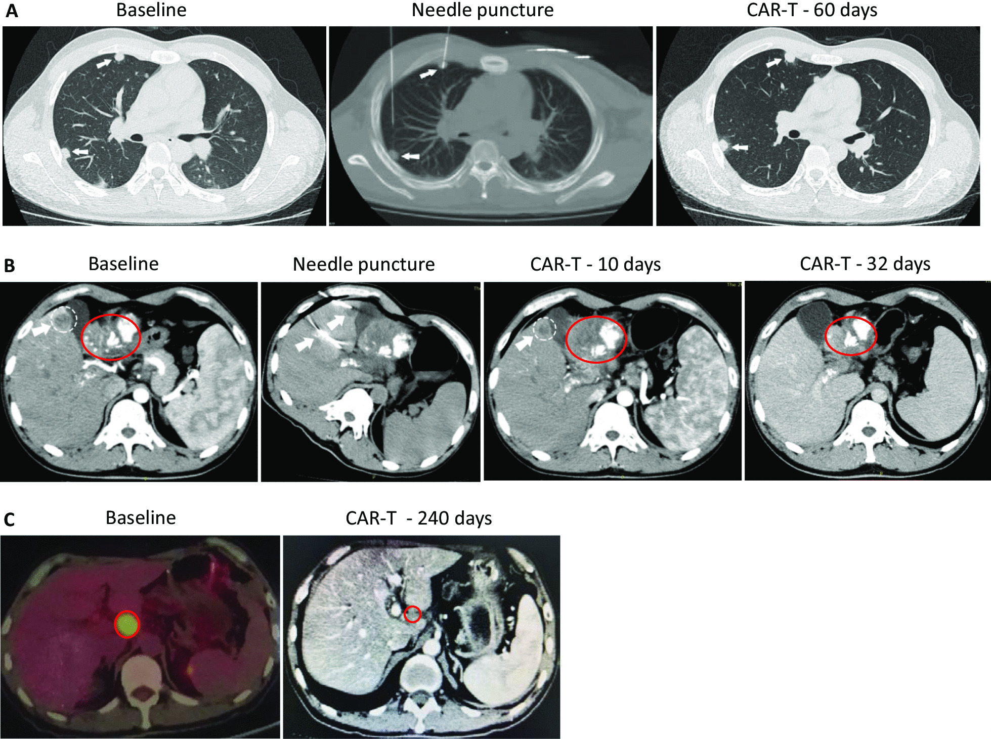
Fig. 2.

7 × 19 CAR-T cells showed remarkable antitumor activity in HCC and PC patients with GPC3 and MSLN expression. For patient GD-G/M-001, three metastatic lesions were chosen to perform fine needles CT-guided intratumor injection of CAR-T cells. a One nodule (0.5 cm) in the right front lung, injected with anti-GPC3 CAR-T cells as a comparison; the second nodule (0.5 cm) in the right back lung, injected with anti-GPC3-7 × 19 CAR-T cells. The results showed that the inhibited growth of anti-GPC3-7 × 19 and anti-GPC3 CAR-T cells at similar levels 60 days after injection. Tumor is indicated by white arrow. b For the same patient GD-G/M-001, a tumor close to the gallbladder (1.7 × 2.0 cm) was fine needle punctured under CT guidance and injected with anti-GPC3-7 × 19 CAR-T cells. 10- and 32-day follow-up CT scans showed a significant decrease in the size of the lesion and finally disappeared, respectively. At the same time, injection of the same CAR-T cells into the portal vein resulted in partial reopening of the previously obstructed portal vein. Metastatic tumor is indicated by white cycle; primary tumor is indicated by red cycle; white arrow points fine needle. c PET-CT and CT scans demonstrated a hepatic hilar lymph node metastasis after primary pancreatic carcinoma surgery in complete response in patient GD-G/M-005 following one intraartery and four intravein administrations of the autologous anti-MSLN-7 × 19 CAR-T cell infusion products. Tumor is indicated by red cycle
