Fig. 2.
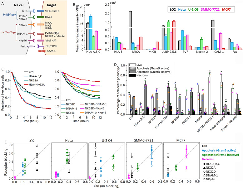
Differential activation of the NK cell inhibitory and activating receptor signaling machinery upon interaction with the five epithelial target cell lines. a Diagram of the major NK cell receptors and their cognate ligands on the target cell. b Comparison of the surface protein expression in the five target cell lines based on the average florescent signal from the flow cytometry analysis. Cell lines were color-coded as indicated. Data were averaged from 3 independent flow cytometry analyses (> 1 × 10⁴ cells in each analysis). The error bars are standard deviations. c Cumulative survival curves of HeLa cells in co-culture with primary NK cells in the presence of single or double blockage of the inhibitory receptor (left panel) or activating receptor (right panel). d Distributions of the live and dead HeLa cells killed by the three distinct cytotoxic modes after 12 h of co-culture with NK cells under the indicated treatment conditions. Data acquired with NK cells from the same donors are denoted with the same color symbols. P value was obtained by Student’s t test comparing the treatment condition with control. *P < 0.001. e Percentage of live cells and cells that died via the three distinct cytotoxic modes under the different receptor-blocking conditions in comparison with those under the control condition. Data are color-coded as indicated. The different treatment conditions are denoted with the indicated symbols. For both d and e, data plotted were averaged from 3 independent imaging experiments and the error bars are standard deviations

