Fig. 3.
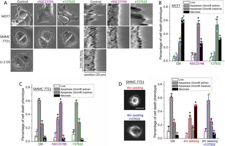
Membrane dynamics of MCF7 and SMMC-7721 cells modulate NK cell cytotoxic modes. a Left panel: phase-contrast images of MCF7, SMMC-7721, and U-2 OS cells under control or treatment with NSC23766 (Rac1 inhibitor) or Y27632 (ROCK inhibitor). The white scale bar is 20 μm. Right-panel: kymographs of a cell edge of MCF7, SMMC-7721, and U-2 OS cells under the indicated treatment conditions. The horizontal axis of the kymographs is the line position across the cell edges, spanning 20 μm, and the vertical axis is the time evolution from 0 to 60 min. b, c Distributions of the cytotoxic response phenotypes of target cells under control or the indicated inhibitor treatment for b MCF7 cells or c SMMC-7721 cells in co-culture with primary NK cells for 12 h. d Phase-contrast images and the response phenotype distribution of SMMC-7721 cells that adhered to the cell culture plate for 4 h (with or without Y27632) and in co-culture with primary NK cells for 12 h in comparison with control cells that adhered for 24 h. Data plotted in a–d were averaged from 3 independent imaging experiments, and the number of cells analyzed for each condition/cell line/experiment ranges from 44 to 149. The error bars are standard deviations. Data acquired with NK cells from the same donors are denoted with the same color symbols in each sub-figure. P value was obtained by Student’s t test comparing the treatment condition with control. *P < 0.001

