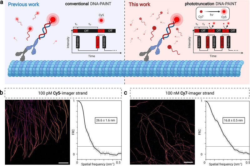
Figure 6.
Cy7-Phototruncation DNA-PAINT. (a) Schematic to apply cyanine phototruncation for conventional and phototruncation DNA-PAINT imaging. (b, c) DNA-PAINT images of immunolabeled microtubules in COS7 cells using conventional Cy5-imager strands (100 pM) and phototruncation Cy7-imager strands (100 nM) and corresponding Fourier ring correlation (FRC) analysis38 estimated spatial resolutions. Measurements were performed in PBS, pH 7.4 containing 500 mM NaCl in the absence (conventional DNA-PAINT) and presence of 10 mM histidine (phototruncation DNA-PAINT) at an irradiation of 0.5 kW cm–2 at 640 nm (τb = bright time, τd = dark time). Scale bars: 5 μm (b, c).

