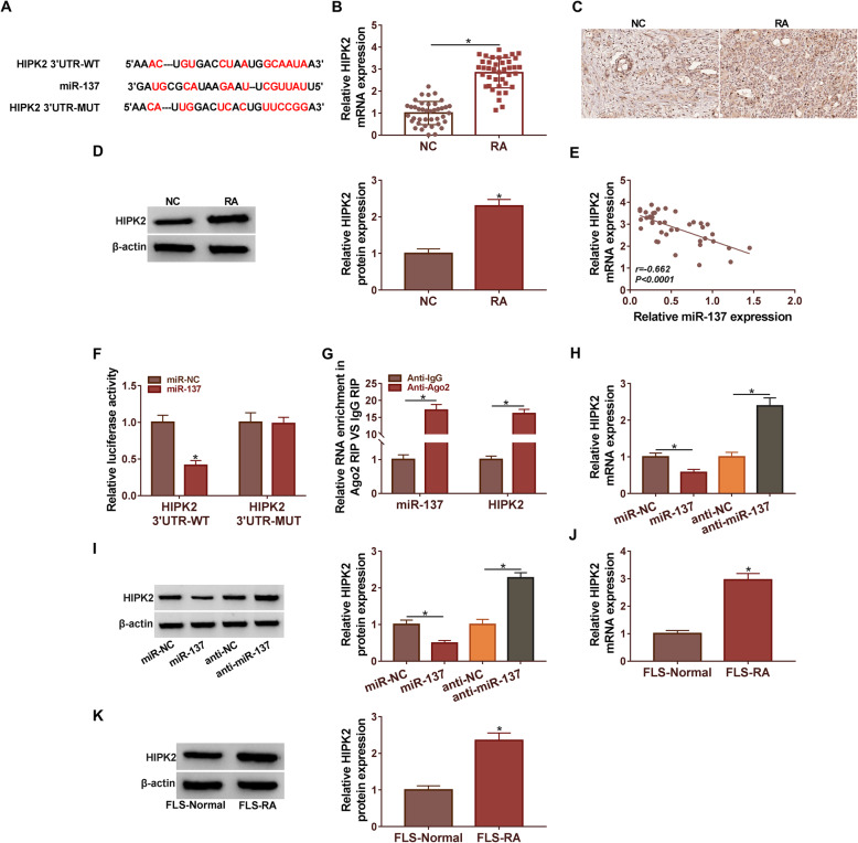
Fig. 5.
MiR-137 targeted HIPK2 in RA cells. (A) The binding site between miR-137 and HIPK2 was analyzed by Starbase. (B and C) The expression of HIPK2 in RA tissues and normal tissues was detected by qRT-PCR and western blot. (D) Pearson’s correlation analysis confirmed that HIPK2 was negatively associated with miR-137 (R = −0.662) in RA tissues. (E) Dual-luciferase reporter assay was used to confirm the relationship between miR-137 and HIPK2. HIPK2 3′UTR-WT: HIPK2 3′UTR wild type, HIPK2 3′UTR-MUT: HIPK2 3′UTR mutant type. (F) RIP assay was used to verify the relationship between HIPK2 and miR-137. (G and H) The expression of HIPK2 in RA cells was detected by qRT-PCR and western blot. (I and J) The expression of HIPK2 in RA cells was detected by qRT-PCR and western blot. *P < 0.05

