Figure 8.
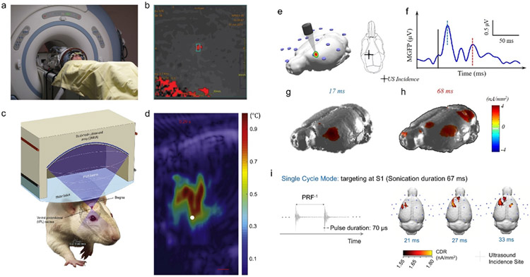
Imaging Guided Transcranial Focused Ultrasound. a)-b) Patient setup with MR guided focused ultrasound, and the thermal spatial mapping for monitoring the focused ultrasound targeting (the panels are adapted from [215]). c)-d) Dual-mode ultrasound array for ultrasound imaging guided focused ultrasound for targeted neuromodulation through thermal effects (the panels are adapted from [217]). e)-h) EEG-based source imaging reconstructing the brain activations in response to low-intensity, non-thermal tFUS with the initial activity at the acoustic administration spot and the observed ancillary activities at other cortical brain areas at a later time. The panels are adapted and revised from [154]. i) The single cycle mode with an ultrasound pulse duration of 70 μs elicited the brain activations at the targeted cortical regions. EEG-source images show cortical activities at 21 ms, 27 ms, and 33 ms after onset of tFUS[224], which is demonstrated to capture the brain dynamics induced by the low-intensity sonication. This panel is adapted from [224].

