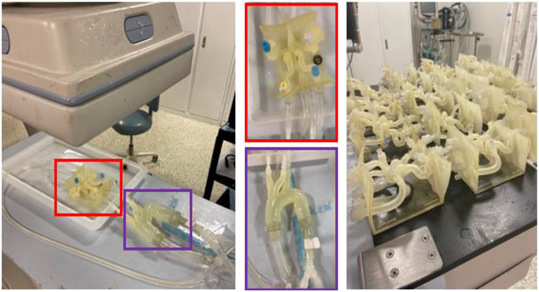
Figure 3:

The 3D printed patient specific Circle of Willis is connected to a flow loop. (Red) The patient specific neurovasculature and (Purple) a standard aortic arch are highlighted. 20 different patient specific models have been printed. A clot is introduced into the proximal MCA and tests the effectiveness of stent retriever thrombectomy with TICI scoring system in patients with absent or robust collaterals of the circle of Willis using a conventional vs. BGC.
