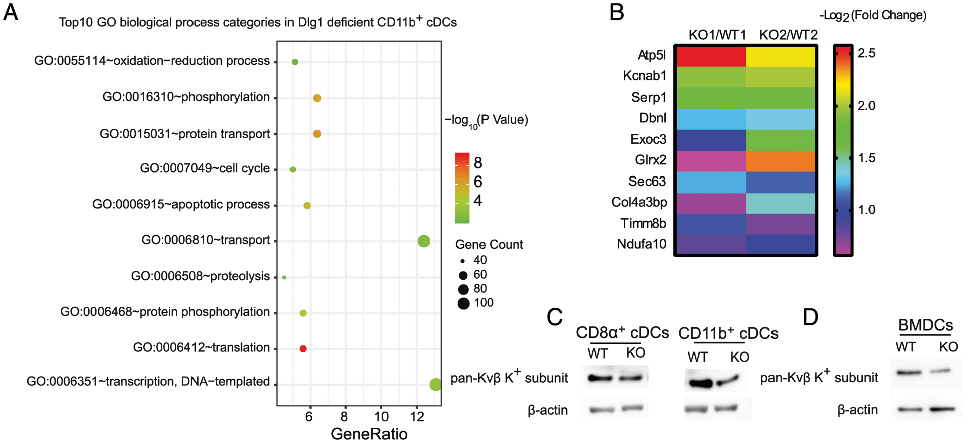
FIGURE 5.

Dlg1 deficiency does not affect TLR signaling and the transcription of proinflammatory cytokines. (A) Top 10 GO BP categories in identified proteins (both upregulated and downregulated proteins). Plot colors represent the statistical significance. GeneRatio (%) represents the proportion of genes in total genes. Plot sizes represent the identified protein numbers in MS. (B) Top 10 downregulated proteins in the transport BP group from Dlg1-KO CD11b+ cDCs. The 10 downregulated proteins were identified from MS data. Each group (KO/WT) indicated one pair of Dlg1fl/flCd11c-Cre-GFP and Cd11c-Cre-GFP mice. Color bar represents protein fold change. (C and D) Expression levels of pan-Kvβ K+ subunit in DCs from Dlg1fl/flCd11c-Cre-GFP mice and Cd11c-Cre-GFP mice, respectively. Whole-cell lysate samples of WT and Dlg1-KO DC subsets were subjected to Western blotting as indicated and probed with anti–pan-Kvβ subunit primary Ab. Loading controls (β-actin) were derived from the same samples. Splenic cDCs (CD11c+CD8α+ or CD11c+CD11b+) in (C) and BMDCs (CD11c+) in (D) are shown. Data are representative of three independent experiments.
