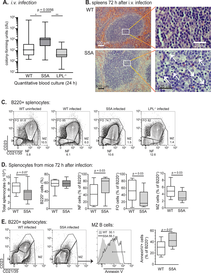
Figure 3. Defective clearance of pneumococcal bacteremia and splenocyte apoptosis in S5A mice.
A. Bacterial burden (cfu) in bloodstream of WT (open, n = 16), S5A (dark gray, n = 17) and LPL−/− (light gray, n = 8) mice 24 h after i.v. challenge with pneumococcus. Kruskal-Wallis ANOVA test, p = 0.0056. Pair-wise comparison of S5A data to WT, p < 0.05; S5A to LPL−/−, p < 0.01 (Dunn’s multiple comparison test). B. Spleens from WT and S5A mice isolated 72 h after i.v. challenge with pneumococcus revealed increased areas of lymphocyte apoptosis in S5A follicles. C. Flow cytometric analysis of B cell subpopulations from uninfected WT, challenged WT, challenged S5A and challenged LPL−/− mice. MZB cells are indicated as CD21/35high/CD23low. D. Quantification of total splenocytes and B cell subpopulations from WT (open, n = 12) and S5A (gray, n = 12) mice 72 h after pneumococcal i.v. challenge. E. Flow cytometric analysis and quantification of Annexin-V labeling of MZB cells derived from WT and S5A mice 72 h after challenge with pneumococcus (i.v.).

