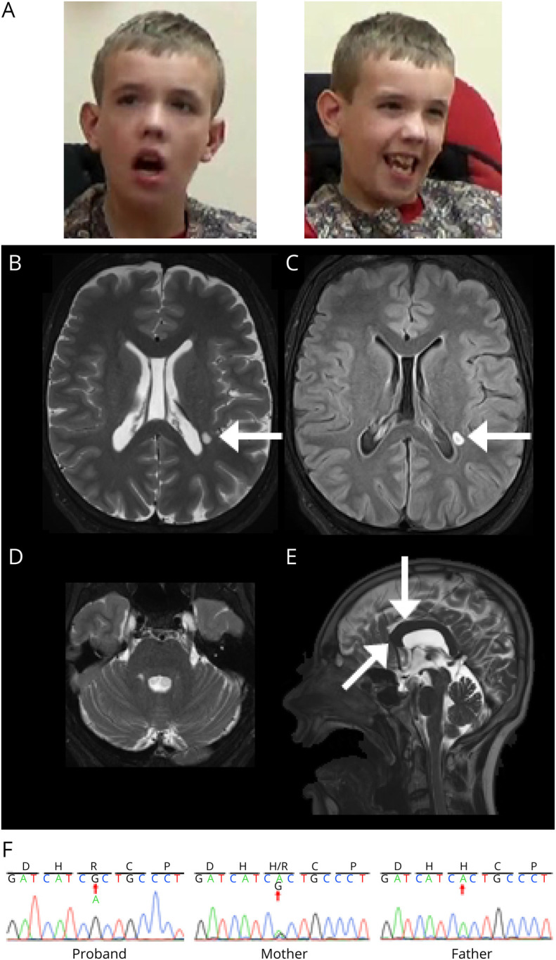
Figure 2. Clinical Features of the Patient With Validated ZDHHC15 Mutation (p.H158R).

(A) Dysmorphic facial features include mild brachycephaly, down-slanting palpebral fissures, large ears, long face, and facial hypotonia. (B–E) Brain MRI at age 17 years. Axial T2 (B) and axial FLAIR (C) images demonstrate mild symmetric widening of the cerebral sulci and a small ovoid focus of T2/FLAIR signal prolongation in the left lateral periatrial white matter (white arrow). Axial FLAIR (D) image at the posterior fossa level demonstrates symmetric widening of the cerebellar folia. Sagittal T2 (E) image at midline shows a smaller-than-expected craniofacial ratio. In addition, the corpus callosum is mildly thickened and foreshortened (white arrow). All these findings have been stable over time on serial MRI. (F) Sanger traces of the proband, mother, and father confirming A to G base change inherited from the mother.
