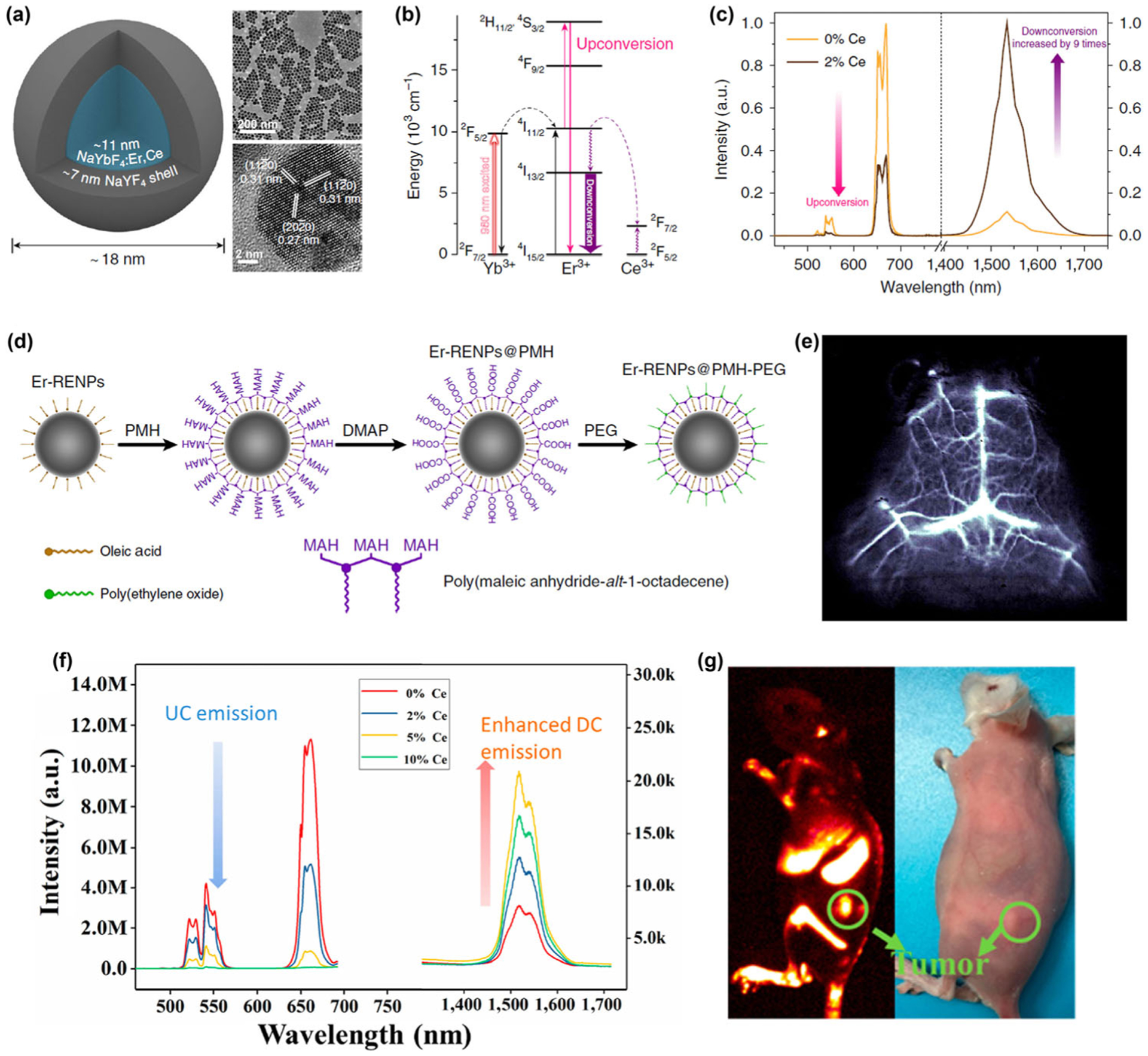
Figure 3.

(a) Schematic design of a NaYbF4:Er,Ce@NaYF4 core-shell nanoparticle (left) with corresponding large scale TEM image (upper right) and HRTEM image (lower right). (b) Simplified energy-level diagrams depicting the energy transfer between Yb3+, Er3+, and Ce3+ ions. (c) Upconversion and down-conversion luminescence spectra of the ErNPs with 0 and 2% Ce3+ doping. (d) Schematic illustration outlining the PMH coating and PEGylation procedure for the ErNPs. (e) Cerebral vascular image (exposure time: 20 ms) in NIR-IIb region by intravenous injection of ErNPs. Reproduced with permission from Ref. [52], © Zhong, Y. T. et al. 2017. (f) Upconversion and NIR-IIb down-conversion luminescence spectra of NaLuF4:Gd/Yb/Er nanorods doped with different contents of Ce3+ (0, 2 mol%, 5 mol%, 10 mol%). (g) Digital photograph (right) of tumor-bearing mouse and in vivo NIR-IIb luminescence imaging (left) of the tumor-bearing mouse (the green circle indicated the tumor site) by intravenous injection of NaLuF4:Gd/Yb/Er/Ce. Reproduced with permission from Ref. [71], © American Chemical Society 2019.
