. 2021 Feb 22;2(2):468–485. doi: 10.1039/d0cb00208a

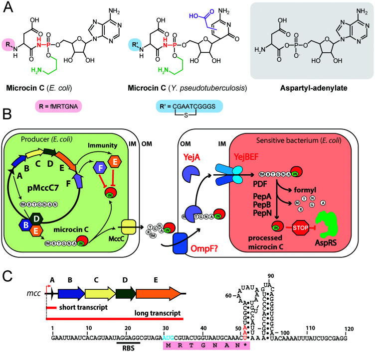
Fig. 4. (A) Chemical structures of peptidyl-nucleotide antibiotics produced by Escherichia coli and Yersinia pseudotuberculosis. The nonhydrolyzable P–N bond is shown in red, the aminopropyl decoration is green, the carboxymethyl attached to the cytosine ring is violet. The peptide part is shown below in single-letter code. Aspartyl-adenylate is shown on the grey background for comparison. (B) Schematic representation of microcin C biosynthesis, transport and processing. PDF – peptide deformylase, OM – outer membrane, IM – inner membrane, AspRS – aspartyl-tRNA synthetase. (C) Regulation of microcin C production by an internal ribosome-dependent transcription terminator located between the mccA and mccB genes. The secondary structure of short mccA transcript is shown.61 Start codon is blue, stop codon is red. RBS – ribosome-binding site.

Fig. 4