. 2021 Feb 22;2(2):468–485. doi: 10.1039/d0cb00208a

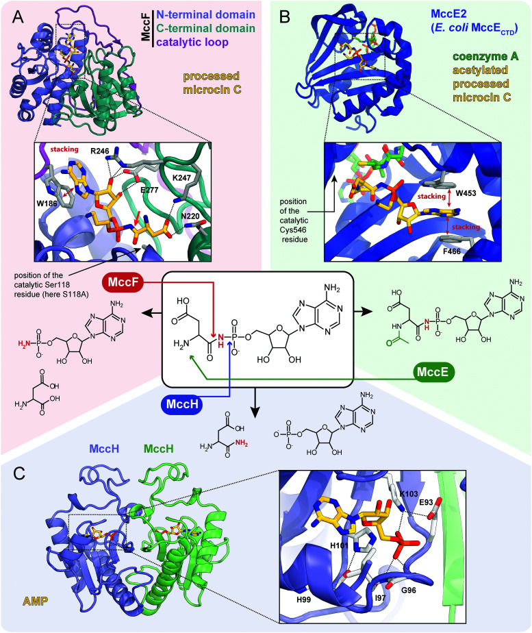
Fig. 7. Proteins providing immunity to microcin C and their mechanisms of action. For each protein the structure, mode of substrate binding, and catalyzed reaction are shown. (A) The structure of E. coli carboxypeptidase MccF catalytical mutant (S118A) in complex with processed microcin C (PDB ID: 3TLC).138 Hydrogen bonds are shown as black dashed lines, π–π stacking between W186 and adenine nucleobase is indicated, big red arrow points to the peptide bond cleaved by MccF. (B) The structure of E. coli acetyltransferase MccE2 (C-terminal domain of MccE, residues 405–589) in complex with coenzyme A and acetylated processed microcin C (PDB ID: 3R9G).93 (C) The theoretical model of dimeric HIT-family hydrolase MccH from Hyalangium minutum in complex with AMP.86.

Fig. 7